


Case #390 - February 2015
 ShareCompartir
A 55-year-old refugee from the Democratic Republic of Congo demonstrated 23.9% eosinophilia with the presence of suspect microfilaria on Wright-Giemsa stained peripheral blood smears. Smears were sent to CDC for confirmation and species identification. Figures A and C show two of the suspect organisms objects observed at 500x magnification with oil; Figures B and C are of the same objects at 1000x magnification with oil. The size of the objects measured 193 micrometers on average. What is your diagnosis? Based on what criteria?
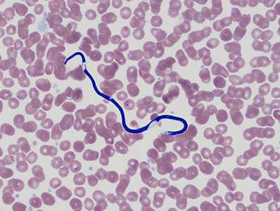
Figure A

Figure B
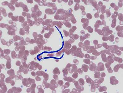
Figure C
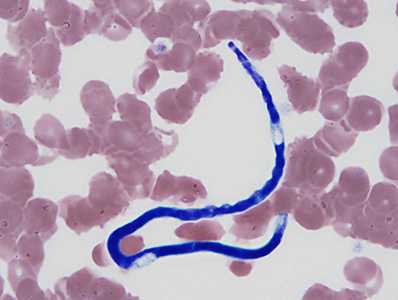
Figure D
Case Answer
This was a case of mansonellosis caused by Mansonella perstans. Diagnostic morphologic features included:
- small, unsheathed microfilariae within the size range for the species (normal range is 190-200 micrometers in length; however shrinkage can occur with processing).
- microfilariae with a compact column of nuclei.
- a blunt tail that contains nuclei to the end of the tail.
In the DRC, M. perstans is similar to ‘Microfilaria’ semiclarum, an enigmatic species of uncertain affinities. Microfilariae of ‘Microfilaria’ semiclarum are larger (198-221 μm long, on average) and have a clear area measuring approximately 40 μm mid-body that is nearly devoid of nuclei, possessing only a few large, scattered nuclei.
More on: mansonellosis
Images presented in the monthly case studies are from specimens submitted for diagnosis or archiving. On rare occasions, clinical histories given may be partly fictitious.
DPDx is an education resource designed for health professionals and laboratory scientists. For an overview including prevention and control visit www.cdc.gov/parasites/.
- Page last reviewed: August 24, 2016
- Page last updated: August 24, 2016
- Content source:
- Global Health – Division of Parasitic Diseases and Malaria
- Notice: Linking to a non-federal site does not constitute an endorsement by HHS, CDC or any of its employees of the sponsors or the information and products presented on the site.
- Maintained By: