


Case #381 - October 2014
 ShareCompartir
A man was seen as an outpatient at a hospital in Atri, Italy. A stool specimen was collected and processed for ova-and-parasite (O&P) testing. Images of suspect parasites were taken and sent to the DPDx Team for diagnostic assistance (Figures A and B). Measurements shown were included in the original telediagnosis submission. No other information was available. What is your diagnosis? Based on what criteria?
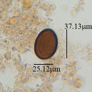
Figure A
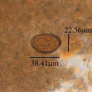
Figure B
Case Answer
The objects shown were the eggs of the trematode Dicrocoelium dendriticum. Diagnostic morphologic features shown in the figures included thick-shelled, dark brown embryonated eggs within the size range for D. dendriticum (35-45 µm long by 20-30 µm wide). Like with many cases involving trematodes, the operculum was not readily visible on either of the eggs. Most infections are light and asymptomatic. However, eggs found in stool can sometimes represent spurious passage and additional specimens should be collected to confirm a true infection.
More on: dicrocoeliasis
Images presented in the monthly case studies are from specimens submitted for diagnosis or archiving. On rare occasions, clinical histories given may be partly fictitious.
DPDx is an education resource designed for health professionals and laboratory scientists. For an overview including prevention and control visit www.cdc.gov/parasites/.
- Page last reviewed: August 24, 2016
- Page last updated: August 24, 2016
- Content source:
- Global Health – Division of Parasitic Diseases and Malaria
- Notice: Linking to a non-federal site does not constitute an endorsement by HHS, CDC or any of its employees of the sponsors or the information and products presented on the site.
- Maintained By: