


Dicrocoeliasis
 ShareCompartir
[Dicrocoelium dendriticum]
Causal Agents
The trematode Dicrocoelium dendriticum, the lanceolate fluke.
Life Cycle
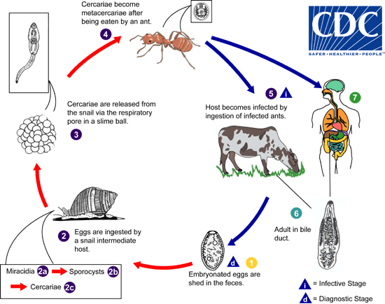
Ruminants are the usual definitive hosts for Dicrocoelium dendricitum, although other herbivorous animals, carnivores, and humans can serve as definitive hosts. Embryonated eggs are shed in feces  . The eggs are ingested by a snail
  . Many species of snail may serve as the first intermediate host, including Zebrina spp. and Cionella spp. When the miracidia hatch
  , they migrate through the gut wall and settle into the adjacent vascular connective tissue, where they become mother sporocysts
  . The sporocysts migrate to the digestive gland where they give rise to several daughter sporocysts. Inside each daughter sporocyst, cercariae are produced
  . The cercariae migrate to the respiration chamber where they are shed in slime ball from the snail
  . After a slime ball is ingested by an ant, the cercariae become free in the intestine and migrate to the hemocoel where they become metacercariae
  . Many ants may serve as the second intermediate host, especially members of the genus, Formica. After an ant is eaten by the definitive host
  , the metacercariae excyst in the small intestine. The worms migrate to the bile duct where they mature into adults
  . Humans can serve as definitive hosts after accidentally ingesting infected ants
  .
.
Geographic Distribution
Europe, northern Asia, America and northern Africa.
Clinical Presentation
Most infections are light and asymptomatic. In heavier infections, symptoms may include cholecystitis, liver abscesses and upper abdominal pain.
Dicrocoelium dendriticum eggs in wet mounts.
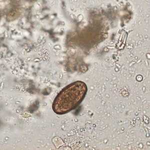
Figure A: Egg of Dicrocoelium dendriticum in an unstained wet mount of stool. Image courtesy of Dr. Juan Cuadros González.
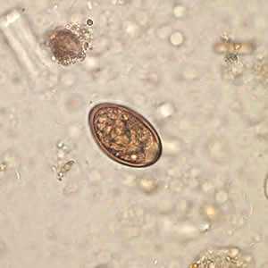
Figure B: Egg of Dicrocoelium dendriticum in an unstained wet mount of stool. Image courtesy of Dr. Juan Cuadros González.
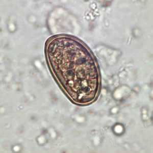
Figure C: Egg of Dicrocoelium dendriticum in an unstained wet mount of stool. Image courtesy of Dr. Juan Cuadros González.
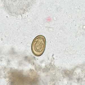
Figure D: Egg of D. dendriticum in an unstained wet mount of stool.
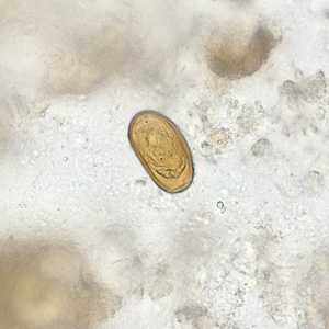
Figure E: Egg of D. dendriticum in an unstained wet mount of stool.
Dicrocoelium dendriticum adults.
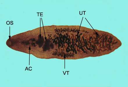
Figure A: Adult of D. dendriticum, stained with carmine. Structures illustrated in this figure include: oral sucker (OS), acetabulum (AC), uterus (UT), testes (TE), and vitelline glands (VT).
Intermediate hosts of Dicrocoelium dendriticum.
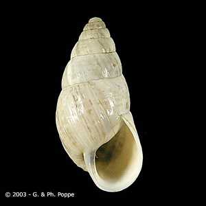
Figure A: Zebrina detrita, a common first intermediate host for D. dendriticum. Image courtesy of Conchology, Inc, Mactan Island, Philippines.
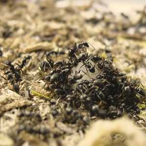
Figure B: Formica fusca, a common second intermediate host for D. dendriticum in Europe. Image courtesy of Sedeer El-Showk.
Laboratory Diagnosis
Microscopic identification of eggs in the stool or duodenal fluid. If eggs are found only in stool, it could represent spurious passage following the ingestion of infected animal liver. Additional specimens should be collected to confirm a true infection.
Treatment Information
In humans, the diagnosis of dicrocoeliasis is typically spurious based on parasite eggs that are passed in the feces after consumption of infected animal liver. True infections are rare. Infection has been treated successfully with praziquantel, 25 mg/kg three times orally daily for 2 days. Praziquantel is not approved for treatment of children less than 4 years old. Niclosamide is effective but is not available in the United States.
DPDx is an education resource designed for health professionals and laboratory scientists. For an overview including prevention and control visit www.cdc.gov/parasites/.
- Page last reviewed: May 3, 2016
- Page last updated: May 3, 2016
- Content source:
- Global Health – Division of Parasitic Diseases and Malaria
- Notice: Linking to a non-federal site does not constitute an endorsement by HHS, CDC or any of its employees of the sponsors or the information and products presented on the site.
- Maintained By: