


Case #366 - February 2014
 ShareCompartir
A 28-year-old male from Algeria had complaints of right upper quadrant (RUQ) abdominal pain. As part of his work up at a medical facility, imaging studies revealed a complex solid and cystic lesion in the posterior right hepatic dome. Hepatic cyst fluid was aspirated and submitted for ova-and-parasite (O&P) testing. Figures A-D show what was observed on a wet-mount made from the sediment of the fluid. Figures A and C were captured at 100x magnifications; Figures B and D at 200x magnification. What is your diagnosis? Based on what criteria?
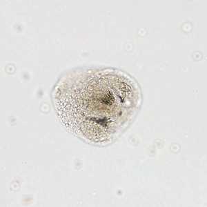
Figure A
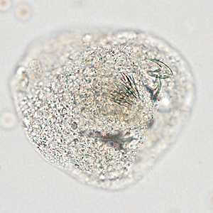
Figure B
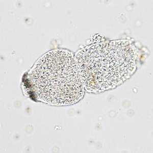
Figure C
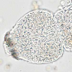
Figure D
Case Answer
This was a case of echinococcosis caused by Echinococcus granulosus. The images showed individual protoscoleces with characteristic suckers and refractile hooks. The presence of a single cyst was also compatible with E. granulosus. Serologic tests can be very helpful in the diagnosis or confirmation of echinococcosis, and may be used before invasive methods.
More on echinococcosis
Images presented in the monthly case studies are from specimens submitted for diagnosis or archiving. On rare occasions, clinical histories given may be partly fictitious.
DPDx is an education resource designed for health professionals and laboratory scientists. For an overview including prevention and control visit www.cdc.gov/parasites/.
- Page last reviewed: August 24, 2016
- Page last updated: August 24, 2016
- Content source:
- Global Health – Division of Parasitic Diseases and Malaria
- Notice: Linking to a non-federal site does not constitute an endorsement by HHS, CDC or any of its employees of the sponsors or the information and products presented on the site.
- Maintained By: