


Case #360 - November 2013
 ShareCompartir
A county health department in Kansas routinely collects stool specimens as part of their refugee screening program. Stool is collected in 10% formalin and zinc-PVA (Zn-PVA) and forwarded to the state health laboratory for ova-and-parasite (O&P) examination. Images of any suspicious objects and organisms are captured and sent to the DPDx Team whenever there is a need for confirmatory diagnosis. Figures A-D show three such objects observed on a trichrome-stained slide of stool preserved in Zn-PVA. A scale bar was included in Figure B to show the average size of the objects. The objects in Figures C and D represent the same structure in two different focal planes. What is your diagnosis? Based on what criteria?
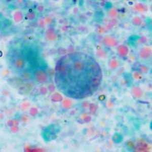
Figure A
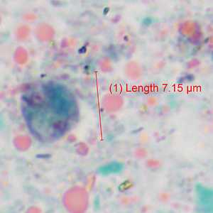
Figure B
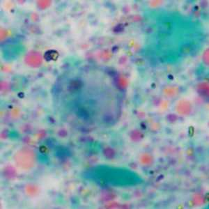
Figure C
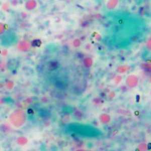
Figure D
Case Answer
This was a case of Dientamoeba fragilis infection. All three organisms demonstrated the binucleate form of the trophozoite and were within the size range (5-15 micrometers) of the species. The nuclei lack peripheral chromatin and possess a fragmented karyosome. The trophozoite in Figure C shows the large central vacuole sometimes observed in D. fragilis.
More on:Dientamoeba fragilis
Images presented in the monthly case studies are from specimens submitted for diagnosis or archiving. On rare occasions, clinical histories given may be partly fictitious.
DPDx is an education resource designed for health professionals and laboratory scientists. For an overview including prevention and control visit www.cdc.gov/parasites/.
- Page last reviewed: August 24, 2016
- Page last updated: August 24, 2016
- Content source:
- Global Health – Division of Parasitic Diseases and Malaria
- Notice: Linking to a non-federal site does not constitute an endorsement by HHS, CDC or any of its employees of the sponsors or the information and products presented on the site.
- Maintained By: