


Case #352 - July, 2013
 ShareCompartir
A 60-year-old Canadian presented to his health care provider with recurrent subcutaneous swellings in his posterior chest. The only travel outside of Canada reported in the last year was to Florida. Biopsy specimens were taken and sent to Pathology for routine histological work-up. Examination of slides stained with hematoxylin and eosin (H&E) revealed a coiled nematode approximately 60 micrometers in diameter (Figures A-D). What is your diagnosis? Based on what criteria? What other testing, if any, would you recommend?
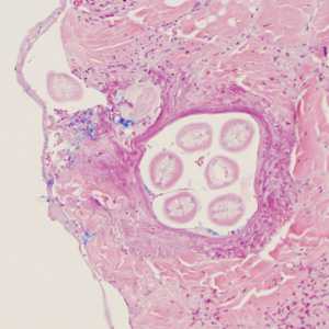
Figure A
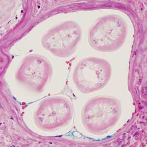
Figure B
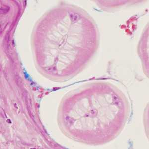
Figure C
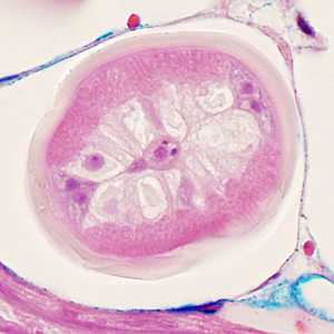
Figure D
Case Answer
This was a case of zoonotic onchocerciasis caused by a nematode in the genus Onchocerca. Diagnostic morphologic features included:
- a relatively thick, smooth cuticle.
- muscle cells tall, but few in number (no more than four per quadrant); basal, fibrillar contractile portion rather diffuse in nature.
- simple, nucleated intestine (best seen in Figure D).
- lack of internal cuticular ridges.
- lack of external lateral alae.
The sections of worm available for study were all cross sections, and so some of the diagnostic features associated with this genus (e.g. external cuticular ridges) were not evident and could not be evaluated. Preliminary molecular analysis also supported the diagnosis of a nematode in the genus Onchocerca. At the time of this writing, a species-level identification has yet to be made.
More on: Onchocerciasis
This case and images were kindly provided by the Mayo Clinic, Rochester, MN and St. Martha’s Regional Hospital, Antigonish, Nova Scotia, Canada.
Images presented in the monthly case studies are from specimens submitted for diagnosis or archiving. On rare occasions, clinical histories given may be partly fictitious.
DPDx is an education resource designed for health professionals and laboratory scientists. For an overview including prevention and control visit www.cdc.gov/parasites/.
- Page last reviewed: August 24, 2016
- Page last updated: August 24, 2016
- Content source:
- Global Health – Division of Parasitic Diseases and Malaria
- Notice: Linking to a non-federal site does not constitute an endorsement by HHS, CDC or any of its employees of the sponsors or the information and products presented on the site.
- Maintained By: