


Case #334 - October, 2012
 ShareCompartir
During the laboratory component of the DPDx Diagnostic Parasitology Workshop on Intestinal Parasites, a participant made an observation on a trichrome-stained fecal smear and wanted to share with the class. The images in Figures A and B were captured at 1000x oil magnification. The two images show the same object in different focal planes. What is your diagnosis? Based on what criteria?
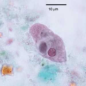
Figure A
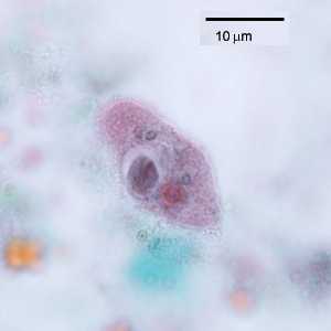
Figure B
Case Answer
The figures showed a trophozoite of Entamoeba coli with an ingested cyst of Giardia duodenalis. Diagnostic features included:
- an amoeba trophozoite with coarse cytoplasm and within the size range for E. coli.
- a single nucleus with an eccentric karyosome.
- a large vacuole that contained a cyst within the size range for G. duodenalis demonstrating intracytoplasmic fibrils, two nuclei.
More on: Giardiasis: Entamoeba coli
Images presented in the monthly case studies are from specimens submitted for diagnosis or archiving. On rare occasions, clinical histories given may be partly fictitious.
DPDx is an education resource designed for health professionals and laboratory scientists. For an overview including prevention and control visit www.cdc.gov/parasites/.
- Page last reviewed: August 24, 2016
- Page last updated: August 24, 2016
- Content source:
- Global Health – Division of Parasitic Diseases and Malaria
- Notice: Linking to a non-federal site does not constitute an endorsement by HHS, CDC or any of its employees of the sponsors or the information and products presented on the site.
- Maintained By: