


Case #333 - October, 2012
 ShareCompartir
A 68-year-old woman underwent a routine screening mammogram, which revealed calcifications in the right breast. Biopsy specimens were collected and sent to Pathology for histological work-up. Figures A–D show what was observed at 20x, 100x, 200x, and 400x magnification respectively by the attending pathologist on one of the slides stained with hematoxylin and eosin (H&E). What is your diagnosis? Based on what criteria?
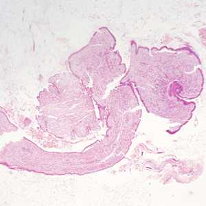
Figure A
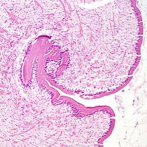
Figure B
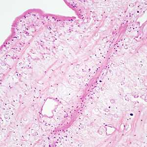
Figure C
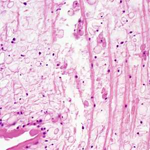
Figure D
Case Answer
This was a case of sparganosis caused by the third-stage plerocercoid larva (sparganum) of a cestode in the genus, Spirometra. Diagnostic features include:
- a thick tegument.
- presence of calcareous corpuscles.
- absence of defined protoscoleces.
More on: Sparganosis
Images presented in the monthly case studies are from specimens submitted for diagnosis or archiving. On rare occasions, clinical histories given may be partly fictitious.
DPDx is an education resource designed for health professionals and laboratory scientists. For an overview including prevention and control visit www.cdc.gov/parasites/.
- Page last reviewed: August 24, 2016
- Page last updated: August 24, 2016
- Content source:
- Global Health – Division of Parasitic Diseases and Malaria
- Notice: Linking to a non-federal site does not constitute an endorsement by HHS, CDC or any of its employees of the sponsors or the information and products presented on the site.
- Maintained By: