


Case #331 - September, 2012
 ShareCompartir
A 35-year-old woman, who had emigrated from Azerbaijan five years previous, presented to her health care provider with abdominal pain. A CT scan revealed multiple pancreatic cysts. The patient noted that she was diagnosed with pancreatic cysts while still living in Azerbaijan. The cysts were aspirated and the fluid was sent to Pathology for histological work-up. Figures A-D show what was observed on a slide made from the aspirate, stained with trichrome. The objects of interest measured 12-15 micrometers on average. What is your diagnosis? Based on what criteria??
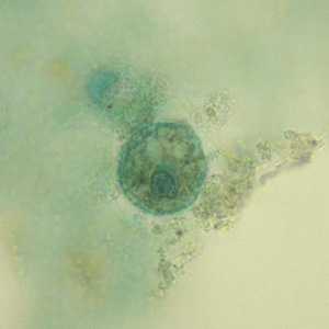
Figure A
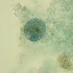
Figure B
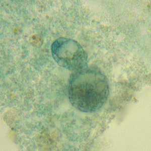
Figure C
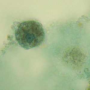
Figure D
Case Answer
This was a case of amebiasis caused by Entamoeba histolytica. Figures A, B, and D showed trophozoites containing a single nucleus with fine, evenly-distributed peripheral chromatin and a discrete central karyosome. The nucleus was not visible in the focal plane shown in Figure C. Although there were no morphologic criteria separating the trophozoites from the similar E. dispar, the location in the pancreas indicates an invasive infection and justifies the species-level identification.
More on: Amebiasis
Images presented in the monthly case studies are from specimens submitted for diagnosis or archiving. On rare occasions, clinical histories given may be partly fictitious.
DPDx is an education resource designed for health professionals and laboratory scientists. For an overview including prevention and control visit www.cdc.gov/parasites/.
- Page last reviewed: August 24, 2016
- Page last updated: August 24, 2016
- Content source:
- Global Health – Division of Parasitic Diseases and Malaria
- Notice: Linking to a non-federal site does not constitute an endorsement by HHS, CDC or any of its employees of the sponsors or the information and products presented on the site.
- Maintained By: