


Case #329 - August, 2012
 ShareCompartir
A seven-year-old boy had an ova-and-parasite (O&P) stool examination performed as part of a required refugee screening. Laboratorians at the state health department observed what they believed to be a few eggs within the size range of 110-120 micrometers. A trematode infection was suspected and images of the eggs were captured and submitted to DPDx for diagnostic assistance. The DPDx team requested that an aliquot of the concentrate be sent for further examination. Figures A-D show what was observed at 200x magnification. The objects of interest measured 115 x 72 micrometers on average. What is your diagnosis? Based on what criteria?
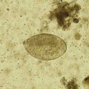
Figure A
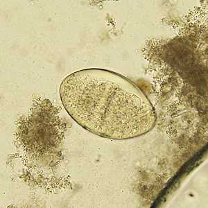
Figure B
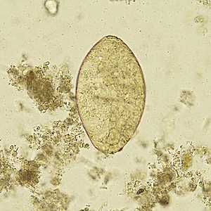
Figure C
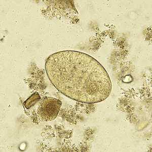
Figure D
Case Answer
This was a case of echinostomiasis caused by a trematode in the genus Echinostoma. Morphologic features shown in the images included:
- eggs within the size range for Echinostoma spp. (80-135 micrometers long by 55-80. micrometers wide), ruling out those of Fasciola and Fasciolopsis, which are both larger (130-150 micrometers long by 60-90 micrometers wide).
- presence of an inconspicuous operculum (Figures A and C).
- thickening of the abopercular end (Figure C).
More on: Echinostomiasis
Images presented in the monthly case studies are from specimens submitted for diagnosis or archiving. On rare occasions, clinical histories given may be partly fictitious.
DPDx is an education resource designed for health professionals and laboratory scientists. For an overview including prevention and control visit www.cdc.gov/parasites/.
- Page last reviewed: August 24, 2016
- Page last updated: August 24, 2016
- Content source:
- Global Health – Division of Parasitic Diseases and Malaria
- Notice: Linking to a non-federal site does not constitute an endorsement by HHS, CDC or any of its employees of the sponsors or the information and products presented on the site.
- Maintained By: