
Echinostomiasis
[Echinostoma spp.]
Causal Agents
Trematodes in the genus, Echinostoma. The genus is worldwide, and about ten species have been recorded in humans, including E. hortense, E. macrorchis, E. revolutum, E. ilocanum and E. perfoliatum. .
Life Cycle
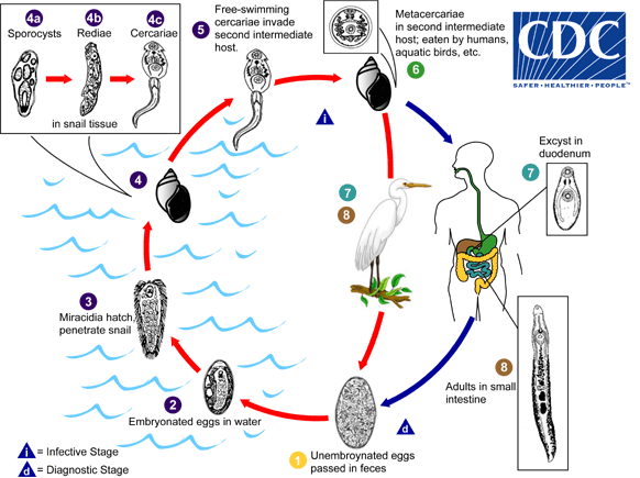
Many animals may serve as definitive hosts for various echinostome species, including aquatic birds, carnivores, rodents and humans. Unembryonated eggs are passed in feces  and develop in the water
 . The miracidium takes on average 10 days to mature before hatching
 and penetrating the first intermediate host, a snail
 . Several genera of snails may serve as the first intermediate host. The intramolluscan stages include a sporocyst
 , one or two generations of rediae
 , and cercariae
 . The cercariae may encyst as metacercariae within the same first intermediate host or leave the host and penetrate a new second intermediate host
 . Depending on the species, several animals may serve as the second intermediate host, including other snails, bivalves, fish, and tadpoles. The definitive host becomes infected after eating infected second intermediate hosts
  . Metacercariae excyst in the duodenum
 and adults reside in the small intestine
 .
.
Geographic Distribution
Worldwide, but human cases are seen most-frequently in southeast Asia and in areas where undercooked or raw freshwater snails, clams and fish are eaten.
Clinical Presentation
Catarrhal inflammation often occurs due to the penetration of the sharp-spined collar into the intestinal mucosa. In heavy infections, nausea, vomiting, diarrhea, fever and abdominal pain may occur.
Echinostoma spp. egg in wet mounts.
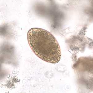
Figure A: Egg of Echinostoma sp. in an unstained wet mount of stool. Image taken at 400x magnification.
Echinostoma spp. adults.
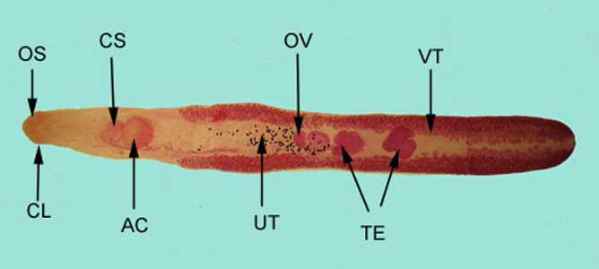
Figure A: Adult of E. revolutum, stained with carmine. Structures illustrated in this figure include: oral sucker (OS), armed collar (CL), cirrus sac (CS), ventral sucker, or acetabulum (AC), uterus containing eggs (UT), ovary (OV), paired testes (TE), and vitelline glands (VT). This species has been recorded from humans in Taiwan.
Echinostoma sp. in tissue, stained with hematoxylin and eosin (H&E).
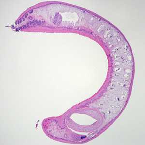
Figure A: Adult Echinostoma removed during a colonoscopy, stained with hematoxylin and eosin (H&E).
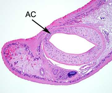
Figure B:Higher magnification of the anterior end of the specimen in Figure A. Notice the acetabulum (ventral sucker, AC).
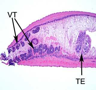
Figure C:Higher magnification of the posterior end of the specimen in Figure A. Notice the vitelline glands (VT) and lobed testes (TE).
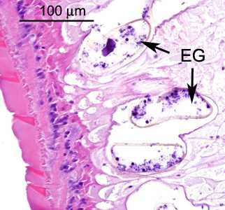
Figure D:Higher magnification of the specimen in Figures A-C. Shown here are eggs (EG) within the size range for Echinostoma spp. (roughly 100 micrometers in length, taking into account they are sections and may not be cut in a perfect horizontal plane).
Intermediate hosts of Echinostoma spp.
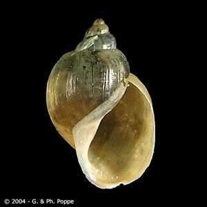
Figure A: Lymnaea sp. This snail genus has been recorded as a second intermediate host for E. malayanum. Image courtesy of Conchology, Inc, Mactan Island, Philippines.
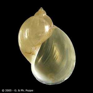
Figure B: Radix sp. This snail genus has been recorded as a first intermediate host for E. hortense and a second intermediate host for E. cinetorchis. Image courtesy of Conchology, Inc, Mactan Island, Philippines.
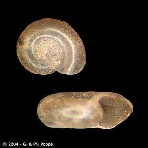
Figure C: Gyraulus sp. This snail genus has been recorded as an intermediate host for E. cinetorchis. Image courtesy of Conchology, Inc, Mactan Island, Philippines.
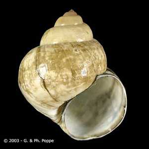
Figure D: Viviparus sp. This snail genus has been recorded as a second intermediate host for E. cinetorchis and E. hortense. Image courtesy of Conchology, Inc, Mactan Island, Philippines.
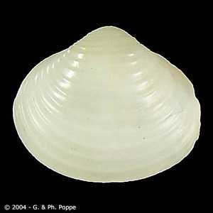
Figure E: Corbicula sp. This bivalve genus has been recorded as a second intermediate host for E. lindoense. Image courtesy of Conchology, Inc, Mactan Island, Philippines.
Laboratory Diagnosis
Microscopic identification of eggs in the stool. Because the eggs are large, careful measurements must be taken to avoid confusion with the eggs of Fasciola or Fasciolopsis. Species-level identification cannot be done based on egg morphology and adults are needed for a definitive diagnosis.
Morphology
More on: morphologic comparisons with other intestinal parasites.
Treatment Information
There are reports of successful removal of Echinostoma flukes from the gastrointestinal tract. Praziquantel* is the anthelminthic drug most commonly used for treatment.
* This drug is approved by the FDA, but considered investigational for this purpose.
DPDx is an education resource designed for health professionals and laboratory scientists. For an overview including prevention and control visit www.cdc.gov/parasites/.
- Page last reviewed: May 3, 2016
- Page last updated: May 3, 2016
- Content source:
- Global Health – Division of Parasitic Diseases and Malaria
- Notice: Linking to a non-federal site does not constitute an endorsement by HHS, CDC or any of its employees of the sponsors or the information and products presented on the site.
- Maintained By:


 ShareCompartir