


Case #326 - June, 2012
 ShareCompartir
A 65-year-old female, who lives in northern California, had a colonoscopy performed as part of a routine screening. A foreign body was observed and subsequently removed. The specimen was sectioned, stained with hematoxylin and eosin (H&E), and examined by the attending pathologist. Digital images were captured and sent to the DPDx Team for diagnostic assistance. Figure A was captured at 2x; Figure B at 20x; and Figures C and D at 40x magnification. What is your diagnosis? Based on what criteria?
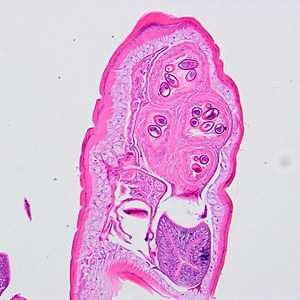
Figure A
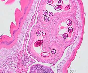
Figure B
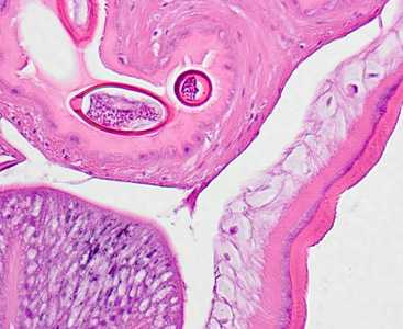
Figure C
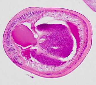
Figure D
Case Answer
This was a case of trichuriasis (commonly called whipworm) caused by the nematode Trichuris trichiura. Morphologic diagnostic features shown included:
- a thick, annulated cuticle.
- a nucleated hypodermis (HY, Figure C) and somatic muscle cells (SM, Figure C).
- a bacillary band (BB, Figure D).
- barrel-shaped eggs with polar plugs (best seen in Figure C).
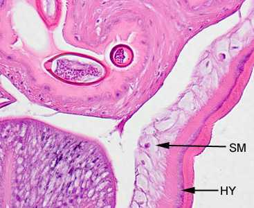
Figure C

Figure D
More on: Trichuriasis
Images presented in the monthly case studies are from specimens submitted for diagnosis or archiving. On rare occasions, clinical histories given may be partly fictitious.
DPDx is an education resource designed for health professionals and laboratory scientists. For an overview including prevention and control visit www.cdc.gov/parasites/.
- Page last reviewed: August 24, 2016
- Page last updated: August 24, 2016
- Content source:
- Global Health – Division of Parasitic Diseases and Malaria
- Notice: Linking to a non-federal site does not constitute an endorsement by HHS, CDC or any of its employees of the sponsors or the information and products presented on the site.
- Maintained By: