


Case #320 - March, 2012
 ShareCompartir
Stool specimens were collected from Haitian refugees as part of a screening program for intestinal parasites. Stool was collected in polyvinyl alcohol (PVA) and 10% formalin and sent to the county public health lab for routine ova-and-parasite (O&P) examination. Figures A-D show what was observed in moderate numbers from one of the patients on a trichrome-stained slide from the PVA-preserved stool. All images were captured at 1000x magnification. What is your diagnosis? Based on what criteria?
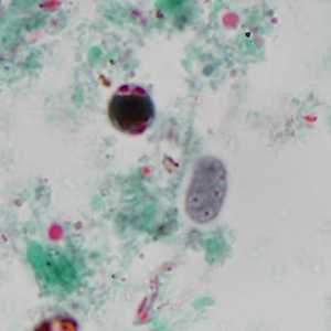
Figure A
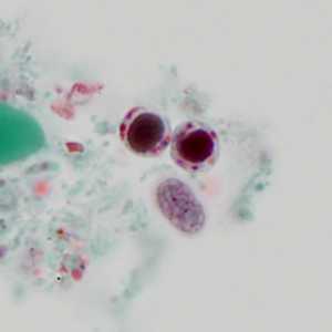
Figure B
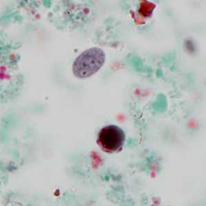
Figure C
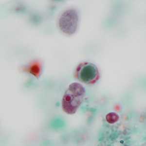
Figure D
Case Answer
This case showed a mix of two intestinal protozoa, Endolimax nana and Blastocystis hominis. Both species were shown in all four images. Diagnostic features included:
- ovoid cysts of E. nana presenting with 2-4 nuclei, each with a discrete karyosome (Figures A-C; there was also a cyst out-of-focus in the upper edge of Figure D).
- trophozoite of E. nana presenting with a single nucleus containing a large, central karyosome and no peripheral chromatin (Figure D).
- cyst-like bodies of B. hominis presenting with a large, central body surrounded by a narrow rim of cytoplasm containing nuclei and inclusion bodies (Figures A-D).
More on: Blastocystis hominis; Endolimax nana
Images presented in the monthly case studies are from specimens submitted for diagnosis or archiving. On rare occasions, clinical histories given may be partly fictitious.
DPDx is an education resource designed for health professionals and laboratory scientists. For an overview including prevention and control visit www.cdc.gov/parasites/.
- Page last reviewed: August 24, 2016
- Page last updated: August 24, 2016
- Content source:
- Global Health – Division of Parasitic Diseases and Malaria
- Notice: Linking to a non-federal site does not constitute an endorsement by HHS, CDC or any of its employees of the sponsors or the information and products presented on the site.
- Maintained By: