


Case #317 - February, 2012
 ShareCompartir
A 55-year-old woman sought medical attention for a nodule on the right side of her head. The patient presented with pain on the right side of her head for the past two months. Two weeks prior to seeking medical attention, she felt a painful 0.5 cm lump on her right temple. The nodule was removed surgically and sent to Pathology for histological testing. Stool and blood specimens were also collected, processed, and examined for parasites with negative results. The nodule was sectioned, stained with hematoxylin and eosin (H&E), and examined microscopically. Images were captured and sent to DPDx for diagnostic assistance. Figure A was captured at 40x; Figure B at 100x; Figures C and D at 200x magnification. What is your diagnosis? Based on what criteria?
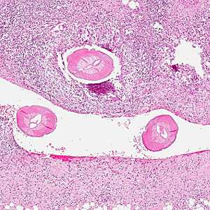
Figure A
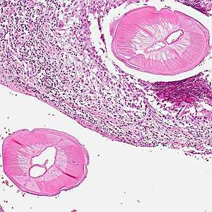
Figure B
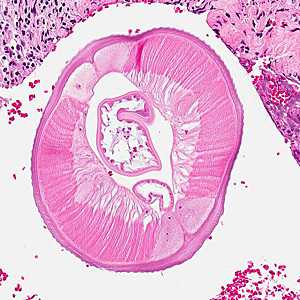
Figure C
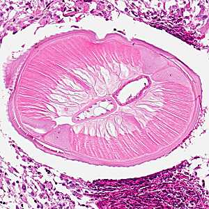
Figure D
Case Answer
This was a case dirofilariasis caused by Dirofilaria sp., most-likely D. repens based on morphologic features and geographic distribution. Morphologic features shown in the images included:
- tall musculature (MU, Figure C).
- large lateral chords (LC, Figure C).
- internal cuticular ridge (IR, Figure C).
- external cuticular ridges (CR, Figure C).
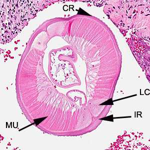
Figure C
More on: Dirofilariasis
This case and images were kindly provided by the National Public Health Surveillance Laboratory, Vilnius, Lithuania.
Images presented in the monthly case studies are from specimens submitted for diagnosis or archiving. On rare occasions, clinical histories given may be partly fictitious.
DPDx is an education resource designed for health professionals and laboratory scientists. For an overview including prevention and control visit www.cdc.gov/parasites/.
- Page last reviewed: August 24, 2016
- Page last updated: August 24, 2016
- Content source:
- Global Health – Division of Parasitic Diseases and Malaria
- Notice: Linking to a non-federal site does not constitute an endorsement by HHS, CDC or any of its employees of the sponsors or the information and products presented on the site.
- Maintained By: