


Case #314 - December, 2011
 ShareCompartir
A 25-year-old man sought medical attention for a nodule on his right hand that had developed about a year after returning from travel to Liberia. The nodule was surgically excised and a tangled mass of thin, white worms was observed within. The worms were preserved in 10% formalin and sent to CDC-DPDx for identification. Figure A shows the mass of worms; Figures B and C show what was observed at 100x after partial clearing in lactophenol. What is your diagnosis? Based on what criteria?
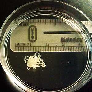
Figure A
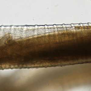
Figure B
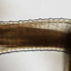
Figure C
Case Answer
This was a case of onchocerciasis caused by Onchocerca volvulus. Morphologic features shown in the images included:
- whitish, thread-like worms that were recovered from a skin nodule (Figure A).
- transverse (annular) thickenings at regular intervals (Figures B and C) along the worms' cuticle.
More on: Onchocerciasis
Images presented in the monthly case studies are from specimens submitted for diagnosis or archiving. On rare occasions, clinical histories given may be partly fictitious.
DPDx is an education resource designed for health professionals and laboratory scientists. For an overview including prevention and control visit www.cdc.gov/parasites/.
- Page last reviewed: August 24, 2016
- Page last updated: August 24, 2016
- Content source:
- Global Health – Division of Parasitic Diseases and Malaria
- Notice: Linking to a non-federal site does not constitute an endorsement by HHS, CDC or any of its employees of the sponsors or the information and products presented on the site.
- Maintained By: