


Case #312 - November, 2011
 ShareCompartir
A 27-year-old patient with multiple myeloma presented with respiratory problems and disseminated lesions over various parts of his body post stem-cell transplant. Bronchial-alveolar lavage (BAL) specimens were collected and sent for pathology work-up. Figures A-D show what was observed on a slide of the BAL specimen stained with Chromotrope 2R. The objects of interest measured 4.0 micrometers in length, on average. What is your diagnosis? Based on what criteria?
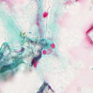
Figure A
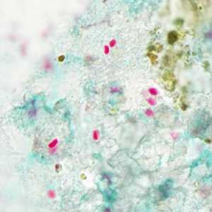
Figure B
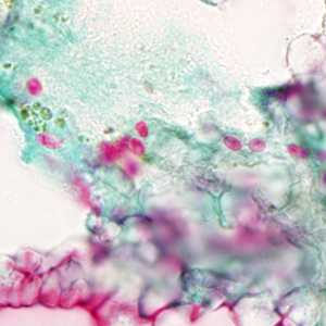
Figure C
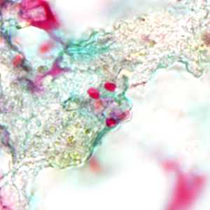
Figure D
Case Answer
This was a case of microsporidiosis. The images showed spores that stained positive with Chromotrope 2R, some of which possessed an equatorial belt. The size was a bit above the range for microsporidia that are known to cause intestinal infections in humans, and diagnostic PCR was negative for Enterocytozoon bieneusi, Encephalitozoon cuniculi, E. hellem, and E. intestinalis. Full length sequencing analysis of the 18S rRNA gene identified the species as Tubulinosema acridophagus.
More on: Microsporidiosis
Images presented in the monthly case studies are from specimens submitted for diagnosis or archiving. On rare occasions, clinical histories given may be partly fictitious.
DPDx is an education resource designed for health professionals and laboratory scientists. For an overview including prevention and control visit www.cdc.gov/parasites/.
- Page last reviewed: August 24, 2016
- Page last updated: August 24, 2016
- Content source:
- Global Health – Division of Parasitic Diseases and Malaria
- Notice: Linking to a non-federal site does not constitute an endorsement by HHS, CDC or any of its employees of the sponsors or the information and products presented on the site.
- Maintained By: