Case #298 - April, 2011
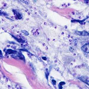
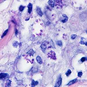
A 41-year-old woman presented to her health care provider with ulcerative lesions on her left ear and neck. Travel history included an excursion along the Amazon River in Brazil four months prior. She also reported numerous insect bites during her trip. The woman was referred to an infectious disease doctor who obtained a biopsy specimen from the largest lesion. The specimen was sent to Pathology where it was sectioned and stained with Giemsa. Figures A and B show what was observed at 1000x magnification. What is your diagnosis? Based on what criteria? What, if any, other testing would you recommend?

Figure A

Figure B
Case Answer
This was a case of leishmaniasis caused by the obligate intracellular protozoan of the genus Leishmania. The diagnosis was based on observing amastigotes in the Giemsa-stained biopsy section. Amastigotes are characterized as being small spherical to ovoid structures approximately 1-5 micrometers in length by 1-2 micrometers wide. In addition, both a nucleus and a kinetoplast must be visualized.
Leishmaniasis in humans can present in two main forms of the disease, cutaneous and visceral. It is important in many cases to isolate the organism in culture for isoenzyme analysis and/or to have molecular testing (PCR) performed on unpreserved material such as biopsy, aspirate, or bone marrow to determine the species of Leishmania. Patient management is dictated by presentation of the infection and species of the parasite. Depending on the species of Leishmania present, if left untreated, cutaneous leishmaniasis can develop into a mucocutaneous form of the disease which can have lasting repercussions for the patient.
More on: Leishmaniasis
Images presented in the monthly case studies are from specimens submitted for diagnosis or archiving. On rare occasions, clinical histories given may be partly fictitious.
DPDx is an education resource designed for health professionals and laboratory scientists. For an overview including prevention and control visit www.cdc.gov/parasites/.
- Page last reviewed: August 24, 2016
- Page last updated: August 24, 2016
- Content source:
- Global Health – Division of Parasitic Diseases and Malaria
- Notice: Linking to a non-federal site does not constitute an endorsement by HHS, CDC or any of its employees of the sponsors or the information and products presented on the site.
- Maintained By:


 ShareCompartir