


Case #294 - February, 2011
 ShareCompartir
A 45-year-old woman, originally from Africa, presented to her primary care provider with diarrhea, nausea, vomiting, weight loss and gastritis. The symptoms occurred approximately three months post kidney transplant. Stool was collected in formalin and polyvinyl alcohol (PVA) for routine ova-and-parasite examination. A wet mount was made from the formalin-preserved stool and observed microscopically by the attending pathologist. Images were captured at 400x magnification and sent to the DPDx Team for diagnostic assistance. Figures A and B show two of the images received for evaluation. Size was not included in the initial inquiry. What is your diagnosis? Based on what criteria?
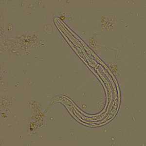
Figure A
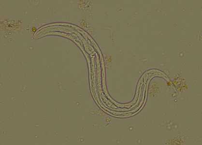
Figure B
Case Answer
This was a case of strongyloidiasis, caused by Strongyloides stercoralis. The images showed first-stage rhabditoid (L1) larvae. Diagnostic morphologic features included:
- a short buccal canal (BC, Figure B).
- a prominent genital primordium (GP, Figure B).
- a rhabditoid esophagus (ES, Figure B).
Although a size was not given, the above features allowed for an accurate identification and to distinguish the organisms from hookworm larvae.

Figure B
More on: Strongyloidiasis
This case and images were kindly provided by Boston Medical Center, Boston, MA.
Images presented in the monthly case studies are from specimens submitted for diagnosis or archiving. On rare occasions, clinical histories given may be partly fictitious.
DPDx is an education resource designed for health professionals and laboratory scientists. For an overview including prevention and control visit www.cdc.gov/parasites/.
- Page last reviewed: August 24, 2016
- Page last updated: August 24, 2016
- Content source:
- Global Health – Division of Parasitic Diseases and Malaria
- Notice: Linking to a non-federal site does not constitute an endorsement by HHS, CDC or any of its employees of the sponsors or the information and products presented on the site.
- Maintained By: