


Case #284 - September, 2010
 ShareCompartir
A 10-year-old immigrant from Haiti was screened for parasites at a refugee screening clinic. Stool specimens were collected in 10% formalin and polyvinyl alcohol and sent to the local state health laboratory for testing. Figures A, C, and E show what was observed at 400x magnification in a wet mount prepared from the formalin-preserved stool. Figures B, D, and F show the same fields, respectively, but in different focal planes. The objects in Figures A and C measured on average 65 micrometers long by 40 micrometers wide. The object in Figure E measured 90 micrometers long by 45 micrometers wide. What is your diagnosis? Based on what criteria?
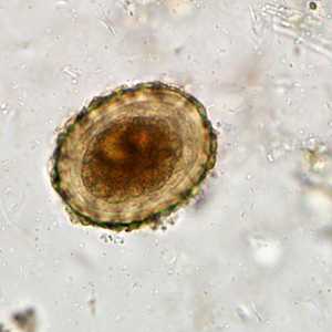
Figure A
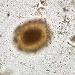
Figure B
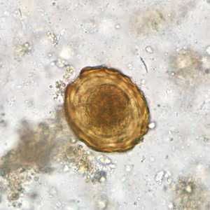
Figure C
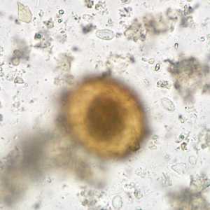
Figure D
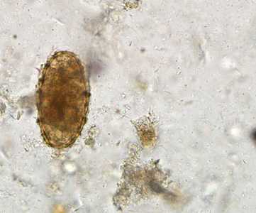
Figure E
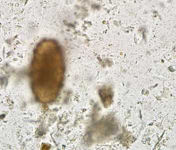
Figure F
Case Answer
This case demonstrated a mixed infection of ascariasis, caused by Ascaris lumbricoides, and giardiasis, caused by Giardia duodenalis. Diagnostic morphologic features included:
- unembryonated, fertile eggs of A. lumbricoides within the size range for the species and demonstrating a thick wall with a mammillated layer (Figures A and C).
- an infertile egg of A. lumbricoides within the size range for the species (Figure E).
- cysts of G. duodenalis (arrows, Figures B, D and F).
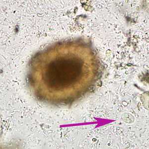
Figure B
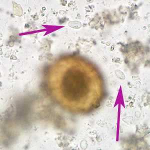
Figure D
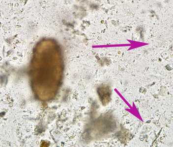
Figure F
More on: Ascariasis: Giardiasis
Images presented in the monthly case studies are from specimens submitted for diagnosis or archiving. On rare occasions, clinical histories given may be partly fictitious.
DPDx is an education resource designed for health professionals and laboratory scientists. For an overview including prevention and control visit www.cdc.gov/parasites/.
- Page last reviewed: August 24, 2016
- Page last updated: August 24, 2016
- Content source:
- Global Health – Division of Parasitic Diseases and Malaria
- Notice: Linking to a non-federal site does not constitute an endorsement by HHS, CDC or any of its employees of the sponsors or the information and products presented on the site.
- Maintained By: