


Case #279 - July, 2010
 ShareCompartir
While examining a malaria-positive smear on a patient from Uganda, microbiologists at the Ontario Agency for Health Protection and Promotion observed what they believed to be a microfilaria. Images were captured and sent via email to the DPDx Team for diagnostic assistance. Figure A shows the object of interest, which measured approximately 200 micrometers in length. Figures B and C show close-ups of the anterior and posterior ends, respectively. What is your diagnosis? Based on what criteria?
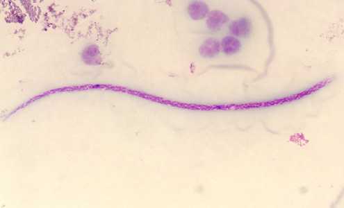
Figure A
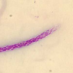
Figure B
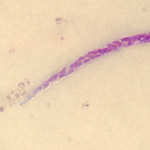
Figure C
Case Answer
This was a case of perstans filariasis caused by Mansonella perstans. Diagnostic morphologic features included:
- an unsheathed microfilaria within the size range for the species (190-200 micrometers in length).
- a compact column of nuclei.
- a blunt tail that contains nuclei to the end of the tail (Figure C).
More on: Mansonellosis
Images presented in the monthly case studies are from specimens submitted for diagnosis or archiving. On rare occasions, clinical histories given may be partly fictitious.
DPDx is an education resource designed for health professionals and laboratory scientists. For an overview including prevention and control visit www.cdc.gov/parasites/.
- Page last reviewed: August 24, 2016
- Page last updated: August 24, 2016
- Content source:
- Global Health – Division of Parasitic Diseases and Malaria
- Notice: Linking to a non-federal site does not constitute an endorsement by HHS, CDC or any of its employees of the sponsors or the information and products presented on the site.
- Maintained By: