


Case #273 - April, 2010
 ShareCompartir
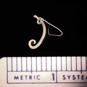
Figure A
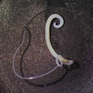
Figure B
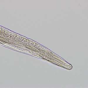
Figure C
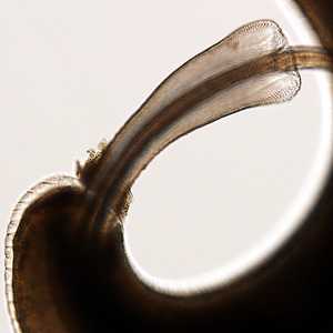
Figure D
Case Answer
This was a case of trichuriasis caused by Trichuris trichiura, also known as the human whipworm. Diagnostic features included a short, thick posterior end and a long, slender, whip-like anterior end (Figures A and B). The coiled posterior end and the presence of a spicule (best seen in Figure D) indicate the specimen was a male. It was unknown at the time whether or not this was a single-worm or unisex worm case, but it should be noted that eggs may not be present during routine ova-and-parasite (O&P) examinations in such cases.
More on: Trichuriasis
Images presented in the monthly case studies are from specimens submitted for diagnosis or archiving. On rare occasions, clinical histories given may be partly fictitious.
DPDx is an education resource designed for health professionals and laboratory scientists. For an overview including prevention and control visit www.cdc.gov/parasites/.
- Page last reviewed: August 24, 2016
- Page last updated: August 24, 2016
- Content source:
- Global Health – Division of Parasitic Diseases and Malaria
- Notice: Linking to a non-federal site does not constitute an endorsement by HHS, CDC or any of its employees of the sponsors or the information and products presented on the site.
- Maintained By: