


Case #263 - November, 2009
 ShareCompartir
A 35-year-old man from Nepal presented at a hospital with recurring cholangitis; the patient had been experiencing symptoms for the previous six months. An MRI revealed two lesions in the liver, measuring approximately 5 centimeters long by 2 centimeters wide. Both lesions had cystic as well as solid components. An endoscopic retrograde cholangiopancreatography (ERCP) was also performed in the common bile duct, revealing flat leaf-shaped objects (Figures A-B). The objects were removed and collected in 10% formalin; they measured on average two centimeters in length (Figure C). One of the specimens was sent to a reference laboratory for identification, where it was stained with carmine (Figure D). What is your diagnosis? Based on what criteria?

Figure A
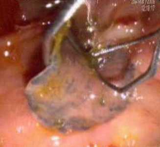
Figure B
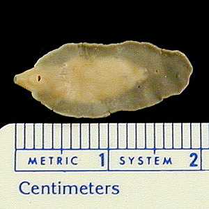
Figure C
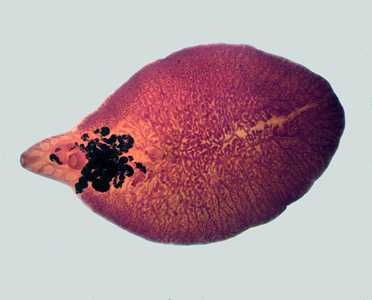
Figure D
Case Answer
This was a case of fascioliasis, caused by the liver fluke, Fasciola hepatica, a large trematode that resides in the bile ducts of the definitive host. Diagnostic features, best seen in the carmine-stained specimen in Figure D, included:
- the presence of oral (OS) and ventral (acetabulum, AC) suckers.
- branching ovaries (OV) and uterus (UT), the latter of which appears dark in carmine-stained specimens.
- extensive branching of the intestinal cecum (CE).
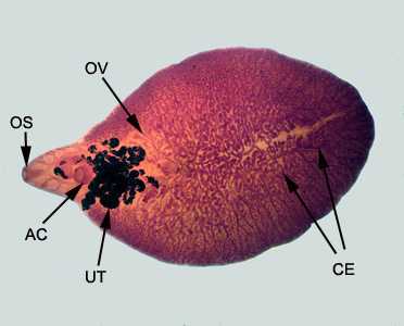
Figure D
More on: Fascioliasis
Images presented in the monthly case studies are from specimens submitted for diagnosis or archiving. On rare occasions, clinical histories given may be partly fictitious.
DPDx is an education resource designed for health professionals and laboratory scientists. For an overview including prevention and control visit www.cdc.gov/parasites/.
- Page last reviewed: August 24, 2016
- Page last updated: August 24, 2016
- Content source:
- Global Health – Division of Parasitic Diseases and Malaria
- Notice: Linking to a non-federal site does not constitute an endorsement by HHS, CDC or any of its employees of the sponsors or the information and products presented on the site.
- Maintained By: