


Case #259 - September, 2009
 ShareCompartir
A four-year-old child was admitted to the hospital for sever abdominal pain mimicking appendicitis. A small section of bowel was removed and sent to the Pathology Department for work-up. A section of tissue was preserved in formalin, sectioned, and stained with hematoxylin and eosin (H&E). Images A-C show what was observed at 100x magnification of slides made from the tissue specimen. In addition to the biopsy, stool was collected for routine ova and parasite (O&P) examination. The object is Figure D, which measured on average 73 micrometers long by 37 micrometers wide, was seen in low numbers in a concentrated wet mount from a formalin-preserved aliquot of the stool. What is your diagnosis? Based on what criteria?
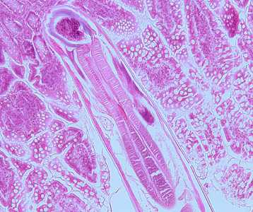
Figure A
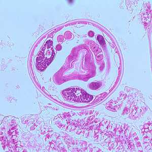
Figure B
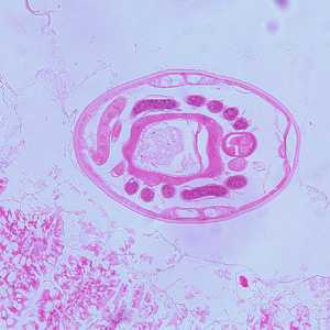
Figure C
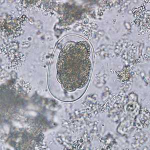
Figure D
Case Answer
This was a case of hookworm infection caused by a member of the genus Ancylostoma or Necator. An examination of intact mouthparts of the adult or male sexual structures is necessary for genus-level identification. Diagnostic features included:
- a longitudinal section of an adult hookworm in tissue demonstrating the oral cavity (OR, Figure A) and a strong, muscled esophagus (ES, Figure A).
- cross-sections of an adult hookworm in tissue demonstrating platymyarian musculature (MU, Figure B), coiled ovaries (OV, Figure B), excretory ducts (ED, Figure B), and an intestine with a brush border (IN, Figure B).
- partially-embryonated eggs in stool with a thin, hyaline shell and within the size range (60-75 micrometers long by 35-40 micrometers wide) for hookworm (Figure D).

Figure A
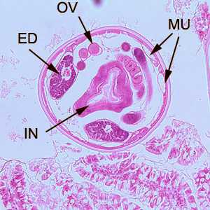
Figure B
More on: Hookworm
Images presented in the monthly case studies are from specimens submitted for diagnosis or archiving. On rare occasions, clinical histories given may be partly fictitious.
DPDx is an education resource designed for health professionals and laboratory scientists. For an overview including prevention and control visit www.cdc.gov/parasites/.
- Page last reviewed: August 24, 2016
- Page last updated: August 24, 2016
- Content source:
- Global Health – Division of Parasitic Diseases and Malaria
- Notice: Linking to a non-federal site does not constitute an endorsement by HHS, CDC or any of its employees of the sponsors or the information and products presented on the site.
- Maintained By: