


Case #258 - August, 2009
 ShareCompartir
A biopsy was performed on a 23-year-old woman with no known travel history, presenting with a perianal ulcer. The specimen was preserved in formalin and sent to a pathology lab for work-up. Figures A and B show what was observed at 500x magnification from a section of the tissue, stained with hematoxylin and eosin (H&E). What is your diagnosis? Based on what criteria?
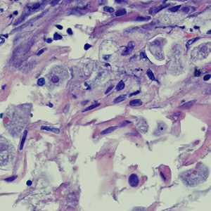
Figure A
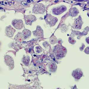
Figure B
Case Answer
This was a case of invasive amebiasis caused by Entamoeba histolytica. Morphologic features seen in the images included:
- the presence of trophozoites in tissue with a single nucleus that contained peripheral chromatin and a central karyosome.
- trophozoites with ingested red blood cells.
More on: Amebiasis
Images presented in the monthly case studies are from specimens submitted for diagnosis or archiving. On rare occasions, clinical histories given may be partly fictitious.
DPDx is an education resource designed for health professionals and laboratory scientists. For an overview including prevention and control visit www.cdc.gov/parasites/.
- Page last reviewed: August 24, 2016
- Page last updated: August 24, 2016
- Content source:
- Global Health – Division of Parasitic Diseases and Malaria
- Notice: Linking to a non-federal site does not constitute an endorsement by HHS, CDC or any of its employees of the sponsors or the information and products presented on the site.
- Maintained By: