


Case #248 - March, 2009
 ShareCompartir
A 36-year-old immigrant from Mozambique presented to his health care provider with heartburn and chronic right abdominal pain. All previous ova and parasite (O&P) examinations were negative. An endoscopy was performed, upon which a worm-like object was observed in the ascending colon near the ileocecum. The suspicious object, measuring approximately 7 mm in length, was removed, collected in 10% formalin, and sent to Pathology for work-up. The object was sectioned, stained with hematoxylin and eosin (H&E) and reviewed by the attending pathologist, who in turn captured the following images and sent them to DPDx for telediagnosis assistance. What is your diagnosis? Based on what criteria?
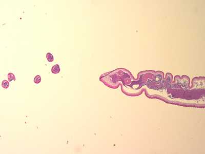
Figure A
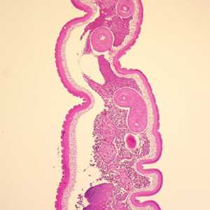
Figure B
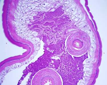
Figure C
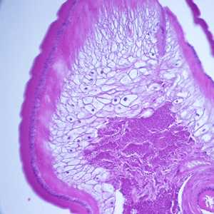
Figure D
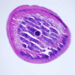
Figure E
Case Answer
This was a case of trichuriasis, caused by Trichuris trichiura. Diagnostic features included:
- the location in the host (colon).
- a marked difference in the size of the anterior and posterior ends of the body (Figure A), with a long, slender anterior end (shown in cross-sections to the left) and a shorter, thicker posterior end (shown as a transverse section to the right).
- a thick cuticle with annulations (arrow, Figure B and CU, Figure D).
- a thin hypodermis (HY, Figure D) containing nuclei.
- the presence of polymyarian muscle cells (PO, Figure D) beneath the hypodermis.
- a thick-muscled cloaca (arrow, Figure C).
- the presence of bacillary bands (BB, Figure E), a stichocyte (ST, Figure E), and stichosome nucleus (SN, Figure E) in the anterior section of the specimen.
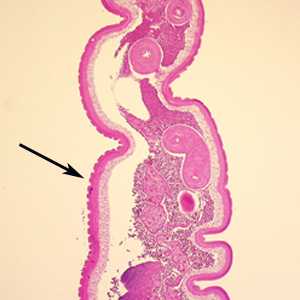
Figure B
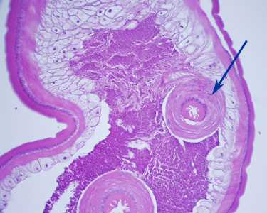
Figure C

Figure D

Figure E
More on: Trichuriasis
Images presented in the monthly case studies are from specimens submitted for diagnosis or archiving. On rare occasions, clinical histories given may be partly fictitious.
This case and images were kindly provided by the Department of Pathology, Cambridge Health Alliance in Cambridge, MA.
DPDx is an education resource designed for health professionals and laboratory scientists. For an overview including prevention and control visit www.cdc.gov/parasites/.
- Page last reviewed: August 24, 2016
- Page last updated: August 24, 2016
- Content source:
- Global Health – Division of Parasitic Diseases and Malaria
- Notice: Linking to a non-federal site does not constitute an endorsement by HHS, CDC or any of its employees of the sponsors or the information and products presented on the site.
- Maintained By: