


Case #240 - November, 2008
 ShareCompartir
A 45-year-old immigrant from Mexico was admitted to the hospital after experiencing headaches, fever, pulmonary symptoms, and adenopathy. Because of his travel history, a blood specimen was collected and sent to Hematology for routine work-up, including blood parasitology. Figures A and B show objects observed on a Giemsa-stained thick smear of the blood specimen. Figures C and D show objects observed on a Giemsa-stained thin smear of the blood specimen. All images were captured at 1000x magnification. The objects in the figures measured on average 180 micrometers in length. What is your diagnosis? Based on what criteria?

Figure A
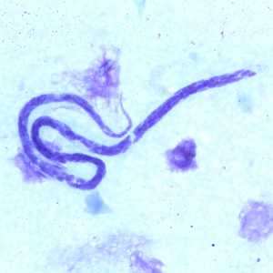
Figure B
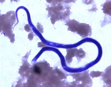
Figure C

Figure D
Case Answer
This was a case of filariasis caused by Mansonella ozzardi. Diagnostic features included:
- microfilariae within the size range (160-205 µm long in stained blood specimens) of the species.
- microfilariae lacking a sheath and containing a compact column of nuclei that terminate well before the end of the tail (arrow, Figure C).
- travel history to Mexico.
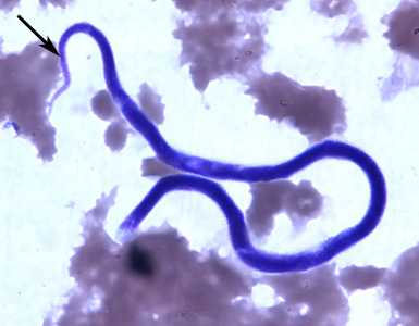
Figure C
More on: Mansonellosis
Images presented in the monthly case studies are from specimens submitted for diagnosis or archiving. On rare occasions, clinical histories given may be partly fictitious.
DPDx is an education resource designed for health professionals and laboratory scientists. For an overview including prevention and control visit www.cdc.gov/parasites/.
- Page last reviewed: August 24, 2016
- Page last updated: August 24, 2016
- Content source:
- Global Health – Division of Parasitic Diseases and Malaria
- Notice: Linking to a non-federal site does not constitute an endorsement by HHS, CDC or any of its employees of the sponsors or the information and products presented on the site.
- Maintained By: