


May - 2008 - Case #227
 ShareCompartir
A 19-year-old male from Louisiana with no known travel history outside the United States presented to his health care provider with a one-month history of headache, fatigue, shortness of breath and weight-loss. A chest radiograph showed significant bilateral pleural effusions with an infiltrate in the left lung. Bronchial alveolar lavage (BAL) and sputum specimens were collected for complete microbiologic and pathologic work-up. Figures A and B show Papanicolaou-stained concentrates from the BAL specimens. Figure A and B were taken at 100x and 1000x oil magnification, respectively. Objects in the figures measured approximately 85-90 micrometers long by 55-60 micrometers wide. What is your diagnosis? Based on what criteria?
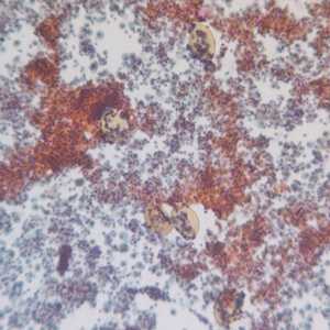
Figure A
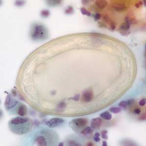
Figure B
Case Answer
The was a case of paragonimiasis caused by the lung fluke, Paragonimus kellicotti. Diagnostic features included:
- the presence of operculated eggs in respiratory specimens (arrows, Figure B point to opercular knobs).
- eggs golden-brown in color, broadest centrally with a thick shell, and within the size range (65-120 micrometers long by 35-70 micrometers wide) of the genus.
- the thickened abopercular end of the eggs.
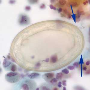
Figure B
Paragonimus kellicotti is of special interest in the United States as this species is endemic to North America. The size range of the eggs of this species and others in the genus (including P. westermani) may overlap, so a species-level identification was not possible in this case based on the characteristics of the eggs alone. A detailed knowledge of the patient’s eating habits and travel history were needed for a complete diagnosis. In this case, the patient consumed raw crayfish, the intermediate host for P. kellicotti, on a dare and had no other travel history that would indicate infection by another Paragonimus species.
More on: Paragonimiasis
These images were kindly contributed by Dr. Gary Procop.
Images presented in the monthly case studies are from specimens submitted for diagnosis or archiving. On rare occasions, clinical histories given may be partly fictitious.
DPDx is an education resource designed for health professionals and laboratory scientists. For an overview including prevention and control visit www.cdc.gov/parasites/.
- Page last reviewed: August 24, 2016
- Page last updated: August 24, 2016
- Content source:
- Global Health – Division of Parasitic Diseases and Malaria
- Notice: Linking to a non-federal site does not constitute an endorsement by HHS, CDC or any of its employees of the sponsors or the information and products presented on the site.
- Maintained By: