


Case #226 - April, 2008
 ShareCompartir
A 32-year-old Latin American man sought medical attention for persistent fever and cough. A sputum specimen was collected for testing. Smears were prepared and stained with Giemsa. The images in Figures A and B show forms that were observed in very low numbers on the smears. Figures A and B were captured at 200x and 1000x oil magnification, respectively. The object measured approximately 490 micrometers in length. What is your diagnosis? Based on what criteria?
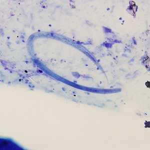
Figure A
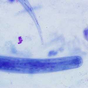
Figure B
Case Answer
This was a case of strongyloidiasis caused by Strongyloides stercoralis. The larva shown in the images was a filariform (infective third-stage form) larva. Diagnostic features included:
- the length of the larva being within the size range for the species.
- the esophagus to intestine ratio, which was roughly 1:1 (Figure A).
- the characteristic notched tail of filariform larvae of S. stercoralis (Figure B).
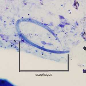
Figure A
More on: Strongyloidiasis
Images presented in the monthly case studies are from specimens submitted for diagnosis or archiving. On rare occasions, clinical histories given may be partly fictitious.
DPDx is an education resource designed for health professionals and laboratory scientists. For an overview including prevention and control visit www.cdc.gov/parasites/.
- Page last reviewed: August 24, 2016
- Page last updated: August 24, 2016
- Content source:
- Global Health – Division of Parasitic Diseases and Malaria
- Notice: Linking to a non-federal site does not constitute an endorsement by HHS, CDC or any of its employees of the sponsors or the information and products presented on the site.
- Maintained By: