


Case #222 - February, 2008
 ShareCompartir
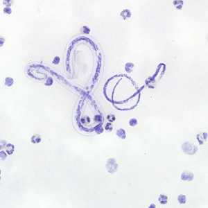
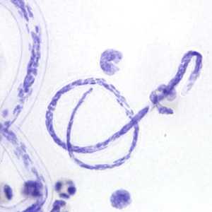
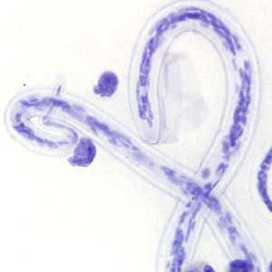
Images were taken from a thick blood smear stained with hematoxylin from an adult male from Cameroon. The following images, A–C were all captured from the same field of view. Figure A was captured at 400x magnification; Figures B and C were captured at 1000x oil magnification. What is your diagnosis? Based on what criteria?
Case Answer
This was a case of filariases caused by both Loa loa and Mansonella perstans. Diagnostic features of the two microfilarie shown in the figures included:
- a larger, more robust microfilaria with a sheath and a tapered tail that contained a single row of nuclei to the very tip of the tail, which are features consistent with Loa loa.
- a smaller microfilaria without a sheath but with a blunt tail filled with nuclei to the tip of the tail, which are features consistent with M. perstans.
The geographic location of the patient (Cameroon) supports this diagnosis as well.
Although size is an important diagnostic feature for separating the various microfilariae found in humans, the above listed features were sufficient to make an accurate diagnosis. One can also use the relative size of white blood cells to estimate the size of the microfilariae.
More on: Loiasis; Mansonellosis
This case and images were kindly contributed by Dr. Juan Cuadros González.
Images presented in the monthly case studies are from specimens submitted for diagnosis or archiving. On rare occasions, clinical histories given may be partly fictitious.
DPDx is an education resource designed for health professionals and laboratory scientists. For an overview including prevention and control visit www.cdc.gov/parasites/.
- Page last reviewed: August 24, 2016
- Page last updated: August 24, 2016
- Content source:
- Global Health – Division of Parasitic Diseases and Malaria
- Notice: Linking to a non-federal site does not constitute an endorsement by HHS, CDC or any of its employees of the sponsors or the information and products presented on the site.
- Maintained By: