


Case #221 - February, 2008
 ShareCompartir
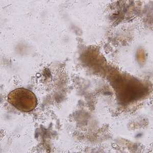
A survey was conducted to determine the prevalence of geohelminth infections in school-age children living in Haiti. The laboratory aspect of the survey consisted of processing stool specimens which were collected in 10% formalin. Per protocol, the processing included performing an FEA (formalin-ethyl acetate) concentration and examination of a wet mount. The following images show what was found in one of the specimens. All images (Figures A–C) were taken at 200x magnification; Figures A and B are of the same field but at different focal planes. The lemon or barrel-shaped objects measured 50-55 micrometers in length. What is your diagnosis? Based on what criteria?


Case Answer
This was a mixed case of trichuriasis and balantidiasis based on the two parasites identifiable in the images: Trichuris trichiura eggs and a Balantidium coli trophozoite. Figures A and B, which were in the same field but different focal planes, showed both parasites. Diagnostic features shown in the images included:
- an egg of T. trichiura in focus in Figure A. Size (given as 50 – 55 micrometers) and shape were consistent with T. trichiura. Polar plugs could also be seen.
- a trophozoite of B. coli (Figure B) showing the cytostome and some cilia.
The large object alongside the T. trichiura egg in Figure C was an artifact (plant cell).
More on: Trichuriasis; Balantidiasis
Images presented in the monthly case studies are from specimens submitted for diagnosis or archiving. On rare occasions, clinical histories given may be partly fictitious.
DPDx is an education resource designed for health professionals and laboratory scientists. For an overview including prevention and control visit www.cdc.gov/parasites/.
- Page last reviewed: August 24, 2016
- Page last updated: August 24, 2016
- Content source:
- Global Health – Division of Parasitic Diseases and Malaria
- Notice: Linking to a non-federal site does not constitute an endorsement by HHS, CDC or any of its employees of the sponsors or the information and products presented on the site.
- Maintained By: