


Case #215 - November, 2007
 ShareCompartir
A 33-year-old Lithuanian woman had swellings in various regions of her body that developed over the last two years. She reported that she worked as a cook, and she also stated she had traveled to Kazakhstan three years ago. A biopsy was taken from a tumor over her right eye lid and was sent to a local pathology laboratory for further examination. Sections were stained with hematoxylin and eosin (H & E) stain. Figures A-D were captured from the sections and emailed to DPDx for diagnostic assistance. What is your diagnosis? Based on what criteria?
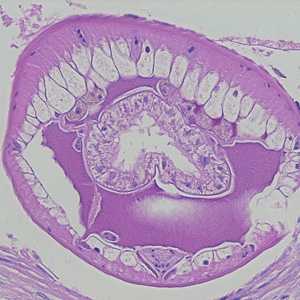
Figure A
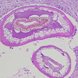
Figure B
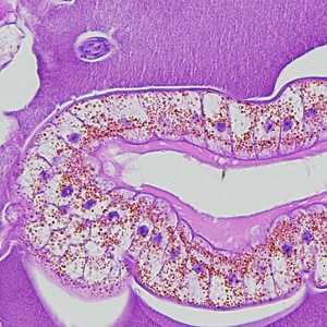
Figure C
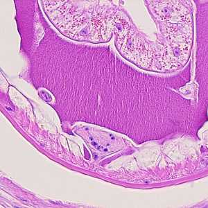
Figure D
Case Answer
This was a case of dioctophymiasis, caused by a nematode in the genus, Dioctophyme (possible D, renale). Diagnostic morphologic features included:
- the presence of a prominent ventral hypodermal chord, containing a U-shaped array of nuclei (black arrow, Figure A).
- the presence of pseudocoelomic membranes (red arrows, Figure A).
- tall, polymyarian musculature.
- nucleated intestinal cells with pigment.
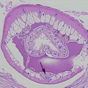
Figure A
More on: Dioctophymiasis
This case was kindly contributed by the Laboratory of Parasitology, National Public Health Research Center in Vilnius, Lithuania.
Images presented in the monthly case studies are from specimens submitted for diagnosis or archiving. On rare occasions, clinical histories given may be partly fictitious.
DPDx is an education resource designed for health professionals and laboratory scientists. For an overview including prevention and control visit www.cdc.gov/parasites/.
- Page last reviewed: August 24, 2016
- Page last updated: August 24, 2016
- Content source:
- Global Health – Division of Parasitic Diseases and Malaria
- Notice: Linking to a non-federal site does not constitute an endorsement by HHS, CDC or any of its employees of the sponsors or the information and products presented on the site.
- Maintained By: