


Dioctophymiasis
 ShareCompartir
[Dioctophyme renale]
Causal Agents
Dioctophyme renale, the giant kidney worm.
Life Cycle
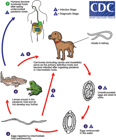
Carnivores, including canids, mustelids and felids, serve as the usual definitive hosts for Dioctophyme renale. However, other mammals, including herbivores and humans, can become infected. Unembryonated eggs are shed in the urine of the definitive host  and L1 larvae develop inside the egg after about a month in water
 . After being eaten by the invertebrate intermediate host (usually annelids, including earthworms), the eggs hatch in the digestive tract and mature into L3 larvae after two molts (usually 2-3 months at 20-30°C). If the intermediate host is eaten by a paratenic host (including fish and amphibians), the L3 larvae encyst and do not develop any further
  . The definitive host then becomes infected after eating a paratenic host housing encysted L3 larvae
  . Definitive hosts may also become infected after directly consuming infected invertebrate intermediate hosts
 . After being ingested by the definitive host, the infective larvae migrate through the gastric wall to the liver, and eventually to the kidney. Worms become adults roughly six months after infecting the definitive host. Humans may also become infected after eating undercooked paratenic hosts
 . Although humans may serve as definitive hosts, often the larvae wind up in subcutaneous nodules and do not develop any further.
Geographic Distribution
Worldwide.
Clinical Presentation
Most of the earlier reports of dioctophymiasis in humans involved the finding of eggs or adult worms in urine. There is one report of a worm rupturing through the body wall (fistula) from an abscessed kidney. There are also a few more recent reports of L3 larvae being found in migratory, subcutaneous nodules.
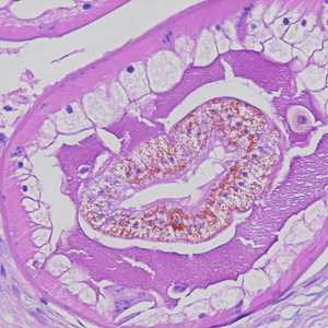
larvae of Dioctotphyme renale in human tissue.
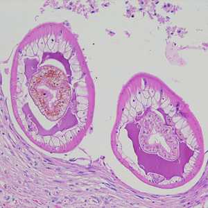
Figure A: Cross-sections of larvae of D. renale in a subcutaneous nodule, stained with hematoxylin and eosin (H&E). Images courtesy of the Laboratory of Parasitology, National Public Health Research Center in Vilnius, Lithuania
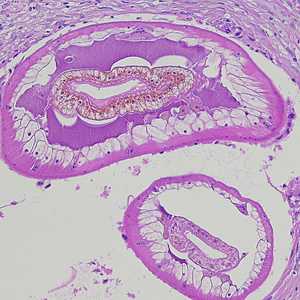
Figure B: Cross-sections of larvae of D. renale in a subcutaneous nodule, stained with hematoxylin and eosin (H&E). Images courtesy of the Laboratory of Parasitology, National Public Health Research Center in Vilnius, Lithuania
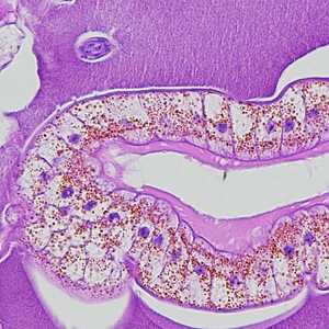
Figure C: Higher-magnification of the specimens shown in Figures A and B, showing a close-up of the characteristic intestine, with cuboidal, uninucleate cells, pigment, and microvilli.
gure D: Higher-magnification of the specimens shown in Figures A and B, showing a close-up of the characteristic intestine, with cuboidal, uninucleate cells, pigment, and microvilli.
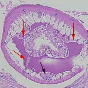
Figure E: Higher-magnification of the specimens shown in Figures A-D. Shown in this image are the tall, polymyarian muscle cells, the characteristic ventral chord with a U-shaped row of nuclei (black arrow), and three pseudocoelomic membranes (red arrows).
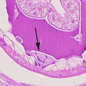
Figure F: Close-up of Figure A, showing the ventral chord (black-arrow).
Eggs of D. renale in animal tissue.
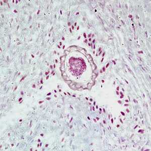
Figure A: Egg of D. renale in the kidney of a mink, stained with hematoxylin and eosin (H&E).
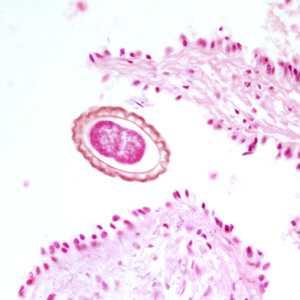
Figure B: Egg of D. renale in the kidney of a mink, stained with hematoxylin and eosin (H&E).
Laboratory Diagnosis
Identification of eggs or adult worms in urine. Adults may also be found during laparotomy or hysterectomy (or necropsy) when in the abdominal cavity. L3 larvae in subcutaneous nodules may be observed in stained tissue sections.
Treatment Information
Due to the rarity of this disease in humans, surgical removal of the worm is the only current recommended treatment.
DPDx is an education resource designed for health professionals and laboratory scientists. For an overview including prevention and control visit www.cdc.gov/parasites/.
- Page last reviewed: May 3, 2016
- Page last updated: May 3, 2016
- Content source:
- Global Health – Division of Parasitic Diseases and Malaria
- Notice: Linking to a non-federal site does not constitute an endorsement by HHS, CDC or any of its employees of the sponsors or the information and products presented on the site.
- Maintained By: