


Case #214 - October, 2007
 ShareCompartir
An 18-year-old woman sought medical attention due to a painful lesion between her toes. She reported travel to Africa. The lesion was excised and sent to CDC for closer examination and identification. The tissue was noted to be fibrous and was carefully dissected to examine what was inside. Figure A shows what was revealed within the tissue; the image was taken with a hand-held digital camera mounted to a dissecting microscope. What is your diagnosis? Based on what criteria?
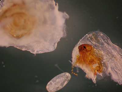
Image A
Case Answer
This was a case of tungiasis caused by the parasitic flea, Tunga penetrans. Travel history and clinical presentation were consistent with tungiasis. Diagnostic features were:
- the presence of a flea in the lesion. The flea had an angular head and did not genal or pronotal combs.
- large, oval egg, indicating that the flea was a gravid female. The size of T. penetrans eggs are approximately 650 micrometers in length.
More on: Tungiasis
Images presented in the monthly case studies are from specimens submitted for diagnosis or archiving. On rare occasions, clinical histories given may be partly fictitious.
DPDx is an education resource designed for health professionals and laboratory scientists. For an overview including prevention and control visit www.cdc.gov/parasites/.
- Page last reviewed: August 24, 2016
- Page last updated: August 24, 2016
- Content source:
- Global Health – Division of Parasitic Diseases and Malaria
- Notice: Linking to a non-federal site does not constitute an endorsement by HHS, CDC or any of its employees of the sponsors or the information and products presented on the site.
- Maintained By: