


Case #193 - December, 2006
 ShareCompartir
A 45-year-old woman had a nodule approximately 1 cm in diameter located near a scar from a previous mastectomy. The patient had traveled to several western and central European countries within the past year. The nodule was removed and tissue sections were stained with hematoxylin and eosin (H & E). Figures A-D show what was seen in the tissue sections. Figure A was taken at 50×, B at 100×, and Figures C and D at 400× magnification respectively. What is your diagnosis? Based on what criteria?
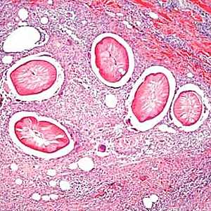
Figure A

Figure B
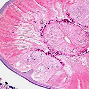
Figure C
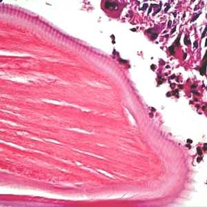
Figure D
Case Answer
This was a case of dirofilariasis caused by Dirofilaria, likely D. repens based on the geographic location and travel history of the patient. The normal host for D. repens is dogs. Diagnostic features were:
- a thick, multi-layered cuticle (Figure C, green bracket) with longitudinal ridges (Figure D, green arrow).
- a prominent internal lateral ridge and large lateral chords (Figure C, black and blue arrows respectively).
- well-developed, polymyarian muscle cells (Figures C and D, yellow bracket).
- Lack of paired uterine tubes indicates that this was probably a male worm.
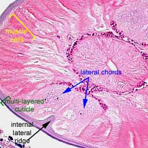
Figure C

Figure D
More on: Dirofilariasis
This case and images were kindly contributed by Dr. Truus Derks.
Images presented in the monthly case studies are from specimens submitted for diagnosis or archiving. On rare occasions, clinical histories given may be partly fictitious.
DPDx is an education resource designed for health professionals and laboratory scientists. For an overview including prevention and control visit www.cdc.gov/parasites/.
- Page last reviewed: August 24, 2016
- Page last updated: August 24, 2016
- Content source:
- Global Health – Division of Parasitic Diseases and Malaria
- Notice: Linking to a non-federal site does not constitute an endorsement by HHS, CDC or any of its employees of the sponsors or the information and products presented on the site.
- Maintained By: