


Case #192 - November, 2006
 ShareCompartir
A request for confirmation of an identification was submitted to DPDx Telediagnosis Assistance. Figures A and B were captured from a hematoxylin and eosin (H & E) stained histological section of a liver abscess found during an autopsy. The patient had a travel history to Sri Lanka. What is your diagnosis? Based on what criteria?

Figure A
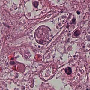
Figure B
Case Answer
This was a case of invasive extraintestinal amebiasis caused Entamoeba histolytica. Diagnostic features were:
- the size of the nuclei in the trophozoites, which is smaller than those seen in macrophages. The ratio for macrophages is generally 1:4 to 1:6, whereas the E. histolytica/dispar ratio is 1:10 to 1:12.
- nuclei with even peripheral chromatin. In Figure B a distinct central karyosome was present (black arrow). In pathological preparations all morphologic features may not be visualized due angle of the cuts.
- location of these organisms in liver tissue, which is consistent with invasive extraintestinal amebiasis and rules-out the morphologically-similar, E. dispar.
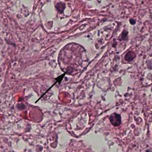
Figure B
In addition to finding organisms in tissue, antigen detection or molecular testing should be considered for a confirmatory diagnosis.
More on: Entamoeba histolytica
This case was kindly contributed by the Oregon State Public Health Laboratory.
Images presented in the monthly case studies are from specimens submitted for diagnosis or archiving. On rare occasions, clinical histories given may be partly fictitious.
DPDx is an education resource designed for health professionals and laboratory scientists. For an overview including prevention and control visit www.cdc.gov/parasites/.
- Page last reviewed: August 24, 2016
- Page last updated: August 24, 2016
- Content source:
- Global Health – Division of Parasitic Diseases and Malaria
- Notice: Linking to a non-federal site does not constitute an endorsement by HHS, CDC or any of its employees of the sponsors or the information and products presented on the site.
- Maintained By: