


Case #185 - August, 2006
 ShareCompartir
A worm measuring approximately 11 mm in length was sent to CDC for identification by a laboratory in the Southeastern United States. The following images were obtained by placing the worm on a 1″ × 3″ glass slide and gently “floating” a 24 × 30 mm glass coverslip on top of it with water. Figure A shows the anterior end of the worm. Figures B and C (a digital zoom of B) show the posterior end of the worm. All images were captured at 100× magnification. Based on the images, identification at the genus level, as well as determination of whether the worm is male or female, is possible. What is your diagnosis? Based on what criteria?
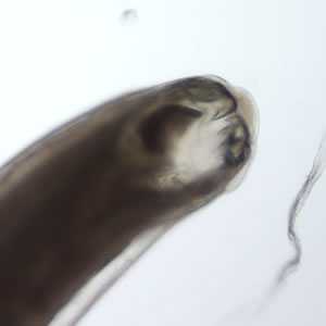
Figure A
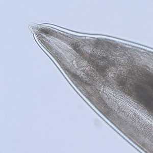
Figure B

Figure C
Case Answer
The worm shown in this case was an adult female Ancylostoma sp. Diagnostic features were:
- the presence of teeth in a large buccal capsule. Necator americanus has cutting plates. Ancylostoma caninum has three pairs of teeth and A. duodenale has two pairs. It was difficult to determine exactly how many pairs were present in this specimen.
- a small spine can be seen at the posterior end in Figure B, and more clearly in Figure C. This feature is seen in female A. caninum.
More on: Hookworm
Images presented in the monthly case studies are from specimens submitted for diagnosis or archiving. On rare occasions, clinical histories given may be partly fictitious.
DPDx is an education resource designed for health professionals and laboratory scientists. For an overview including prevention and control visit www.cdc.gov/parasites/.
- Page last reviewed: August 24, 2016
- Page last updated: August 24, 2016
- Content source:
- Global Health – Division of Parasitic Diseases and Malaria
- Notice: Linking to a non-federal site does not constitute an endorsement by HHS, CDC or any of its employees of the sponsors or the information and products presented on the site.
- Maintained By: