


Case #178 - April, 2006
 ShareCompartir
A 29-year-old man went to a local health clinic with complaints of intermittent diarrhea for a few months. He did not report any other symptoms but said he had difficulty sleeping sometimes. A stool specimen was collected and preserved in both 10% formalin and polyvinyl-alcohol (PVA). An ova and parasite (O & P) examination was performed. Figure A shows what was seen on a concentrated wet mount preparation, and Figure B shows what was seen on a wet mount stained with iodine. Figures C and D show what was seen on a trichrome stained smear made from the PVA preserved specimen. Objects measured 7 to 10 µm in diameter. What is your diagnosis? Based on what criteria?
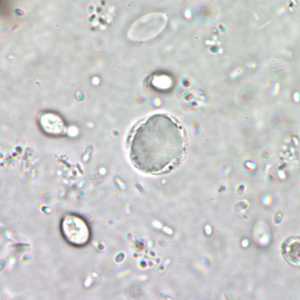
Figure A
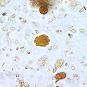
Figure B

Figure C
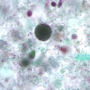
Figure D
Case Answer
The organisms shown in the images are cyst-like forms of Blastocystis hominis. Diagnostic features were:
- the size of the organisms, which was within the range for B. hominis (normally 8 to 10 micrometers in diameter, but can be 5 to 30 micrometers).
- the presence of small nuclei within the peripheral ring of cytoplasm. In trichrome stained preparations, these nuclei often appear red.
- the presence of a large, centrally located vacuole or central body. In trichrome stained preparations, these bodies appear green to dark purple in color.
More on: Blastocystis hominis
Images presented in the monthly case studies are from specimens submitted for diagnosis or archiving. On rare occasions, clinical histories given may be partly fictitious.
DPDx is an education resource designed for health professionals and laboratory scientists. For an overview including prevention and control visit www.cdc.gov/parasites/.
- Page last reviewed: August 24, 2016
- Page last updated: August 24, 2016
- Content source:
- Global Health – Division of Parasitic Diseases and Malaria
- Notice: Linking to a non-federal site does not constitute an endorsement by HHS, CDC or any of its employees of the sponsors or the information and products presented on the site.
- Maintained By: