


Case #159 - July, 2005
 ShareCompartir
A 25-year-old woman had a few mildly scaly lesions on her forehead. She saw her physician and initially it was thought that she had a fungal lesion. Figure A-C show what was seen in skin scrapings from the lesion. The object shown was approximately .25mm long. What is your diagnosis? Based on what criteria?
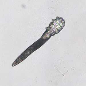
Figure A
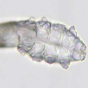
Figure B
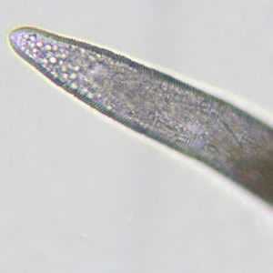
Figure C
Case Answer
The images showed a follicle mite (Demodex sp.). There are two species of Demodex that are associated with humans, D. folliculorum, which lives in the hair follicles and D. brevis, which lives in the sebaceous glands, although there are many species that infest wild and domestic animals. The two species are similar in appearance. Diagnostic features included:
- the size, which was in range for Demodex spp. (0.1 mm to 0.4 mm).
- an elongated body that was somewhat cigar-shaped (the body of D. brevis may be somewhat shorter).
- eight very short, segmented legs.
In general, these mites are harmless, although in some instances they may cause irritations. More images of Demodex can be seen here, on the scabies pages. Please note, Demodex is shown with scabies as a differential diagnosis, especially when examining tissue sections. Demodex spp. are not causative agents of scabies in humans.
More on: Scabies
This case was kindly contributed by Dr. CSBR Prasad, Vindhya Clinic and Diagnostic Lab, India.
Images presented in the monthly case studies are from specimens submitted for diagnosis or archiving. On rare occasions, clinical histories given may be partly fictitious.
DPDx is an education resource designed for health professionals and laboratory scientists. For an overview including prevention and control visit www.cdc.gov/parasites/.
- Page last reviewed: August 24, 2016
- Page last updated: August 24, 2016
- Content source:
- Global Health – Division of Parasitic Diseases and Malaria
- Notice: Linking to a non-federal site does not constitute an endorsement by HHS, CDC or any of its employees of the sponsors or the information and products presented on the site.
- Maintained By: