


Case #158 - June, 2005
 ShareCompartir
A 42-year-old man complained of diarrhea for 2 weeks following an international assignment that included travel to countries in Southeast Asia and the Indian subcontinent. He explained to his physician that his symptoms began shortly before he returned to the United States. An ova and parasites (O & P) examination was ordered, and Figures A-D show what was observed. Figures A and B are from a wet mount stained with Lugol’s iodine, and Figures C and D are from a trichrome stained fecal smear. The objects ranged in size from 20-25 micrometers. What is your diagnosis? Based on what criteria?
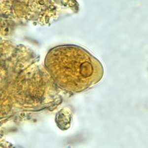
Figure A
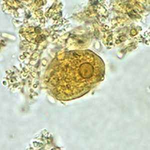
Figure B
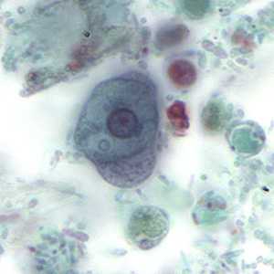
Figure C
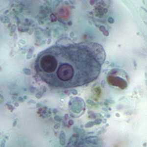
Figure D
Case Answer
This was a case of amebiasis caused by Entamoeba histolytica. Blastocystis hominis was also observed in Figures C and D. Diagnostic features were:
- the size of the organisms, which was within the range trophozoites of E. histolytica/E. dispar (10 to 60 micrometers).
- peripheral chromatin in the nuclei which was evenly distributed
- the karyosome seen in the nuclei, which was centrally located and small. This helps to distinguish from E. coli.
- the cytoplasm, which appeared fine and granular in Figures A and B; Figures C and D appear less so but this may be an effect of less than optimal preservation. The cytoplasm in the trophozoites in Figure C and D does appear fine and granular, especially in the pseudopodia.
- the presence of a pseudopod (Figures A, C and D), a feature more common with, but not exclusive to, E. histolytica/E. dispar.
- a trophozoite with an ingested red blood cell (erythrophagocytosis; red arrow, Figure D), which is a way to distinguish E. histolytica from E. dispar (see image below, with red arrow pointing to the ingested red blood cell).
In addition to seeing ingested red blood cells in trophozoites, molecular tools such as PCR can also be used to make a confirmatory diagnosis of amebiasis. Also shown in the images is Blastocystis hominis (black arrows, Figure D). Diagnostic features of B. hominis included the appearance of a central body, surrounded by a thin rim of cytoplasm containing up to six nuclei. The size of the objects was also within range for B. hominis (5-30 micrometers).
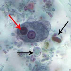
Figure A
More on: Amebiasis: Blastocystis hominis
Images presented in the monthly case studies are from specimens submitted for diagnosis or archiving. On rare occasions, clinical histories given may be partly fictitious.
DPDx is an education resource designed for health professionals and laboratory scientists. For an overview including prevention and control visit www.cdc.gov/parasites/.
- Page last reviewed: August 24, 2016
- Page last updated: August 24, 2016
- Content source:
- Global Health – Division of Parasitic Diseases and Malaria
- Notice: Linking to a non-federal site does not constitute an endorsement by HHS, CDC or any of its employees of the sponsors or the information and products presented on the site.
- Maintained By: