


Case #154 - April, 2005
 ShareCompartir
A 49-year-old male immigrant from Mexico was seen at a local medical facility specializing in neural disorders for frequent headaches and occasional seizures. Figures A and B show what was observed on a hematoxylin and eosin (H & E) stained section of lesions detected in the right frontal lobe of his brain. Figure A was taken at 40× magnification and Figure B was taken at 100× magnification. What is your diagnosis? Based on what criteria?
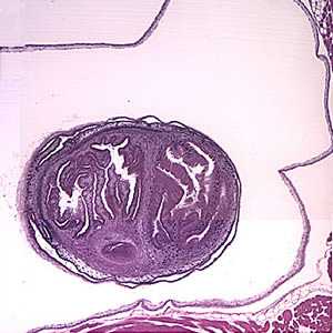
Figure A
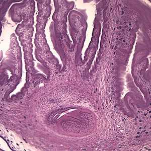
Figure B
Case Answer
This was a case of cysticercosis caused by the larval stage of Taenia solium. Diagnostic features were:
- a single protoscolex (yellow circle, Figure A) visible within the bladder wall (blue arrows, Figure A). Racemose cysticerci are characterized by proliferating, lobulated cysts without a scolex which are usually found in the ventricular system and subarachnoid space, and, rarely, in the cerebral parenchyma.
- the extensive folding of the spiral canal and one sucker of the protoscolex (black arrow, Figure B).
- the presence of calcareous corpuscles (green arrows, Figure B).
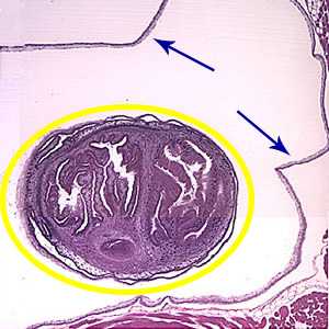
Figure A
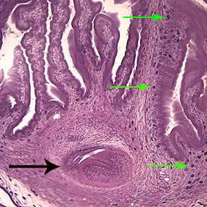
Figure B
Serology for cysticercosis is recommended as a noninvasive laboratory diagnostic procedure prior to treatment that can be very useful in patients suspected of having cysticercosis.
More on: Cysticercosis
Images presented in the monthly case studies are from specimens submitted for diagnosis or archiving. On rare occasions, clinical histories given may be partly fictitious.
DPDx is an education resource designed for health professionals and laboratory scientists. For an overview including prevention and control visit www.cdc.gov/parasites/.
- Page last reviewed: August 24, 2016
- Page last updated: August 24, 2016
- Content source:
- Global Health – Division of Parasitic Diseases and Malaria
- Notice: Linking to a non-federal site does not constitute an endorsement by HHS, CDC or any of its employees of the sponsors or the information and products presented on the site.
- Maintained By: