Upper extremity of humerus
The upper or proximal extremity of the humerus consists of the bone's large rounded head joined to the body by a constricted portion called the neck, and two eminences, the greater and lesser tubercles.
| Upper extremity of humerus | |
|---|---|
 Left humerus. Anterior and posterior views. | |
| Details | |
| Identifiers | |
| Latin | Extremitas proximalis humeri |
| FMA | 23362 |
| Anatomical terms of bone | |
Humeral head
The head (caput humeri), is nearly hemispherical in form. It is directed upward, medialward, and a little backward, and articulates with the glenoid cavity of the scapula to form the glenohumeral joint (shoulder joint). The circumference of its articular surface is slightly constricted and is termed the anatomical neck, in contradistinction to a constriction below the tubercles called the surgical neck which is frequently the seat of fracture. Fracture of the anatomical neck rarely occurs.[1] The diameter of the humeral head is generally larger in men than in women.
Anatomical neck
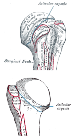
The anatomical neck (collum anatomicum) is obliquely directed, forming an obtuse angle with the body. It is best marked in the lower half of its circumference; in the upper half it is represented by a narrow groove separating the head from the tubercles. It affords attachment to the articular capsule of the shoulder-joint, and is perforated by numerous vascular foramina. Fracture of the anatomical neck rarely occurs.[1]
The anatomical neck of the humerus is an indentation distal to the head of the humerus on which the articular capsule attaches.
Tubercles
The greater tubercle (tuberculum majus; greater tuberosity) is situated lateral to the head and lesser tubercle, and just lateral to the anatomical neck. Its upper surface is rounded and marked by three flat impressions: the highest of these gives insertion to the supraspinatus muscle; the middle to the infraspinatus muscle; the lowest one, and the body of the bone for about 2.5 cm. below it, to the teres minor muscle. The lateral surface of the greater tubercle is convex, rough, and continuous with the lateral surface of the body.[1]
The lesser tuberosity or lesser tubercle (tuberculum minus; lesser tuberosity), although smaller, is more prominent than the greater: it is situated in front, and is directed medialward and forward. Above and in front it presents an impression for the insertion of the tendon of the subscapularis muscle.[1]
Bicipital groove
The tubercles are separated from each other by a deep groove, the bicipital groove (intertubercular groove), which lodges the long tendon of the biceps brachii muscle and transmits a branch of the anterior humeral circumflex artery to the shoulder-joint. It runs obliquely downward, and ends near the junction of the upper with the middle third of the bone. In the fresh state its upper part is covered with a thin layer of cartilage, lined by a prolongation of the synovial membrane of the shoulder-joint; its lower portion gives insertion to the tendon of the latissimus dorsi muscle. It is deep and narrow above, and becomes shallow and a little broader as it descends. Its lips are called, respectively, the crests of the greater and lesser tubercles (bicipital ridges), and form the upper parts of the anterior and medial borders of the body of the bone.[1]
Surgical neck
The surgical neck is a narrow area distal to the tubercles that is a common site of fracture. It makes contact with the axillary nerve and the posterior humeral circumflex artery.
Additional images
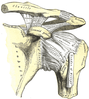 The left shoulder and acromioclavicular joints, and the proper ligaments of the scapula
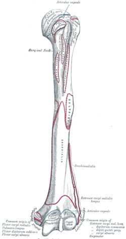 Left humerus. Anterior view.
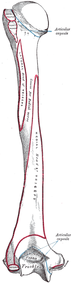 Left humerus. Posterior view.
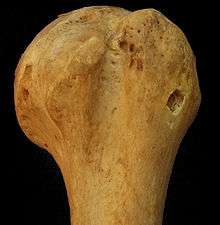 Left humerus. Anteriolateral view.
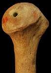 Left humerus. Medial view.
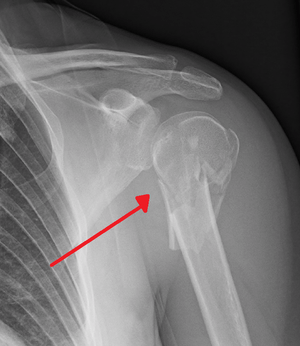 Fracture of the proximal humerus
References
This article incorporates text in the public domain from page 209 of the 20th edition of Gray's Anatomy (1918)
- Gray's Anatomy, see infobox