Olecranon
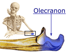
The olecranon /oʊˈlɛkrənɒn/ from the Greek olene meaning elbow and kranon meaning head[1] is the large, thick, curved bony eminence of the ulna, a long bone in the forearm that projects behind the elbow. It forms the most pointed portion of the elbow and is opposite to the cubital fossa or elbow pit. The olecranon serves as a lever for the extensor muscles that straighten the elbow joint.
| Olecranon | |
|---|---|
 | |
| Details | |
| Identifiers | |
| Latin | Olecranon |
| MeSH | D056740 |
| TA | A02.4.06.002 |
| FMA | 39795 |
| Anatomical terms of bone | |
Structure
It is situated at the upper (proximal) end of the ulna, one of the two bones in the forearm. When the hand faces forward (supination) the olecranon faces towards the back (posteriorly).
It is bent forward at the summit so as to present a prominent lip which is received into the olecranon fossa of the humerus in extension of the forearm.
Its base is contracted where it joins the body and the narrowest part of the upper end of the ulna.
Its posterior surface, directed backward, is triangular, smooth, subcutaneous, and covered by a bursa.
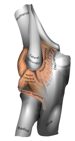
Its superior surface is of quadrilateral form, marked behind by a rough impression for the insertion of the Triceps brachii; and in front, near the margin, by a slight transverse groove for the attachment of part of the posterior ligament of the elbow-joint.
Its anterior surface is smooth, concave, and forms the upper part of the semilunar notch.
Its borders present continuations of the groove on the margin of the superior surface; they serve for the attachment of ligaments, viz., the back part of the ulnar collateral ligament medially, and the posterior ligament laterally.
From the medial border a part of the flexor carpi ulnaris arises; while to the lateral border the anconeus muscle is attached.
Clinical significance
Fractures of the olecranon are common injuries. An olecranon fracture with anterior displacement of the radial head is called a Hume fracture.
Additional images
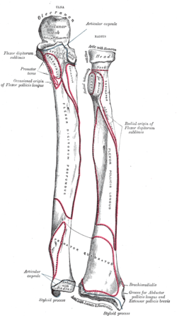 Bones of left forearm. Anterior aspect.
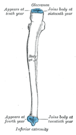 Plan of ossification of the ulna. From three centers.
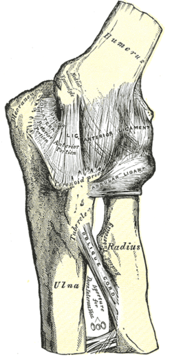 Left elbow-joint, showing anterior and ulnar collateral ligaments.
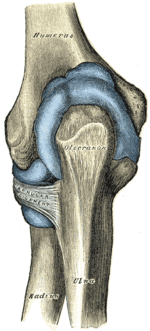 Capsule of elbow-joint (distended). Posterior aspect.
References
This article incorporates text in the public domain from page 214 of the 20th edition of Gray's Anatomy (1918)
- O.D.E. 2nd edition 2005
External links
- radiographsul at The Anatomy Lesson by Wesley Norman (Georgetown University) (xrayelbow)
- "Anatomy diagram: 02240.008-1". Roche Lexicon - illustrated navigator. Elsevier. Archived from the original on 2014-01-01.
| Authority control |
|---|