Type I hypersensitivity
Type I hypersensitivity (or immediate hypersensitivity) is an allergic reaction provoked by re-exposure to a specific type of antigen referred to as an allergen.[1] Type I is distinct from type II, type III and type IV hypersensitivities.
- Not to be confused with Type I Diabetes or Type I of any other disease or reaction.
| Type I hypersensitivity | |
|---|---|
| Other names | Immediate hypersensitivity |
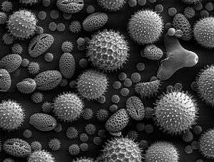 | |
| SEM of miscellaneous plant pollens: Pollens are very common allergens. | |
| Specialty | Immunology |
Exposure may be by ingestion, inhalation, injection, or direct contact.
Pathophysiology
In type 1 hypersensitivity, B-cells are stimulated (by CD4+TH2 cells) to produce IgE antibodies specific to an antigen. The difference between a normal infectious immune response and a type 1 hypersensitivity response is that in type 1 hypersensitivity, the antibody is IgE instead of IgA, IgG, or IgM. During sensitization, the IgE antibodies bind to FcεRI receptors on the surface of tissue mast cells and blood basophils.[2] Mast cells and basophils coated by IgE antibodies are "sensitized". Later exposure to the same allergen cross-links the bound IgE on sensitized cells, resulting in anaphylactic degranulation, which is the immediate and explosive release of pharmacologically active pre-formed mediators from storage granules and concurrent synthesis of inflammatory lipid mediators from arachidonic acid;[3] some of these mediators include histamine, leukotriene (LTC4 and LTD4 and LTB4), and prostaglandin, which act on proteins (e.g., G-protein coupled receptors) located on surrounding tissues.[3] The principal effects of these products are vasodilation and smooth-muscle contraction.
Type 1 hypersensitivity can be further classified into immediate and late-phase reactions. The immediate hypersensitivity reaction occurs minutes after exposure and includes release of vasoactive amines and lipid mediators, whereas the late-phase reaction occurs 2–4 hours after exposure and includes the release of cytokines.[4]
| Vasodilation and increased permeability |
| |
|---|---|---|
| Smooth muscle spasm |
| |
| Leukocyte extravasation |
| |
| Unless otherwise specified, the reference for this table is:[5] | ||
The reaction may be either local or systemic. Symptoms vary from mild irritation to sudden death from anaphylactic shock.
Treatment and prognosis
Treatment usually involves adrenaline (epinephrine), antihistamines, and corticosteroids.
If the entire body is involved, then anaphylaxis can take place, which is an acute, systemic reaction that can prove fatal.
Examples
Some examples:
- Allergic asthma
- Allergic conjunctivitis
- Allergic rhinitis ("hay fever")
- Anaphylaxis
- Angioedema
- Urticaria (hives)
- Eosinophilia
- Penicillin allergy
- Cephalosporin allergy
- Food allergy
- Sweet itch
See also
References
- med/1101 at eMedicine
- "The Adaptive Immune System: Type I Immediate Hypersensitivity". Archived from the original on 2010-07-27. Retrieved 2008-09-22.
- Moon TC, Befus AD, Kulka M (2014). "Mast cell mediators: their differential release and the secretory pathways involved". Front Immunol. 5: 569. doi:10.3389/fimmu.2014.00569. PMC 4231949. PMID 25452755.
This release of pre-formed mediators enables not only rapid anaphylactic reactions and allergic responses but also initiates recruitment of leukocytes to sites of pathogen invasion, activation of innate immune processes, and inflammatory responses (1). ... Two types of degranulation have been described for MC: piecemeal degranulation (PMD) and anaphylactic degranulation (AND) (Figures 1 and 2). Both PMD and AND occur in vivo, ex vivo, and in vitro in MC in human (78–82), mouse (83), and rat (84). PMD is selective release of portions of the granule contents, without granule-to-granule and/or granule-to-plasma membrane fusions. ... In contrast to PMD, AND is the explosive release of granule contents or entire granules to the outside of cells after granule-to-granule and/or granule-to-plasma membrane fusions (Figures 1 and 2). Ultrastructural studies show that AND starts with granule swelling and matrix alteration after appropriate stimulation (e.g., FcεRI-crosslinking).
Figure 1: Mediator release from mast cells
Figure 2: Model of genesis of mast cell secretory granules
Figure 3: Lipid body biogenesis
Table 2: Stimuli-selective mediator release from mast cells - Shiv Pillai MD; Abul K. Abbas MBBS; Andrew Wilson (2011). Cellular and Molecular Immunology: with STUDENT CONSULT Online Access. Philadelphia: Saunders. ISBN 1-4377-1528-1.
- Table 5-2 in:Mitchell, Richard Sheppard; Kumar, Vinay; Abbas, Abul K.; Fausto, Nelson. Robbins Basic Pathology. Philadelphia: Saunders. ISBN 1-4160-2973-7. 8th edition.