Trypanosoma cruzi
Trypanosoma cruzi is a species of parasitic euglenoids. Amongst the protozoa, the trypanosomes characteristically bore tissue in another organism and feed on blood (primarily) and also lymph. This behaviour causes disease or the likelihood of disease that varies with the organism: for example, trypanosomiasis in humans (Chagas disease in South America), dourine and surra in horses, and a brucellosis-like disease in cattle. Parasites need a host body and the haematophagous insect triatomine (descriptions "assassin bug", "cone-nose bug", and "kissing bug") is the major vector in accord with a mechanism of infection. The triatomine likes the nests of vertebrate animals for shelter, where it bites and sucks blood for food. Individual triatomines infected with protozoa from other contact with animals transmit trypanosomes when the triatomine deposits its faeces on the host's skin surface and then bites. Penetration of the infected faeces is further facilitated by the scratching of the bite area by the human or animal host.
| Trypanosoma cruzi | |
|---|---|
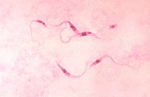 | |
| Trypanosoma cruzi, crithidia | |
| Scientific classification | |
| Phylum: | Euglenozoa |
| Class: | Kinetoplastea |
| Order: | Trypanosomatida |
| Family: | Trypanosomatidae |
| Genus: | Trypanosoma |
| Species: | T. cruzi |
| Binomial name | |
| Trypanosoma cruzi | |
Life cycle
The Trypanosoma cruzi life cycle starts in an animal reservoir, usually mammals, wild or domestic, including humans. A triatomine bug serves as the vector. While taking a blood meal, it ingests T. cruzi. In the triatomine bug (Triatoma infestans) the parasite goes into the epimastigote stage, making it possible to reproduce. After reproducing through binary fission, the epimastigotes move onto the rectal cell wall, where they become infectious. Infectious T. cruzi are called metacyclic trypomastigotes. When the triatomine bug subsequently takes a blood meal from a human, it defecates. The trypomastigotes are in the feces and are capable of swimming into the host's cells using flagella, a characteristic swimming tail dominant in the Euglenoid class of protists.[4]
The trypomastigotes enter the human host through the bite wound or by crossing mucous membranes. The host cells contain macromolecules such as laminin, thrombospondin, heparin sulphate, and fibronectin that cover their surface.[5] These macromolecules are essential for adhesion between parasite and host and for the process of host invasion by the parasite. The trypomastigotes must cross a network of proteins that line the exterior of the host cells in order to make contact and invade the host cells. The molecules and proteins on the cytoskeleton of the cell also bind to the surface of the parasite and initiate host invasion.[5]
Pathophysiology
Trypanosomiasis in humans progresses with the development of the trypanosome into a trypomastigote in the blood and into an amastigote in tissues. The acute form of trypanosomiasis is usually unnoticed, although it may manifest itself as a localized swelling at the site of entry. The chronic form may develop 30 to 40 years after infection and affect internal organs (e.g., the heart, the oesophagus, the colon, and the peripheral nervous system). Affected people may die from heart failure.
Acute cases are treated with nifurtimox and benznidazole, but no effective therapy for chronic cases is currently known.
Cardiac manifestations
Researchers of Chagas’ disease have demonstrated several processes that occur with all cardiomyopathies. The first event is an inflammatory response. Following inflammation, cellular damage occurs. Finally, in the body’s attempt to recover from the cellular damage, fibrosis begins in the cardiac tissue.[6]
Another cardiomyopathy found in nearly all cases of chronic Chagas’ disease is thromboembolic syndrome. Thromboembolism describes thrombosis, the formation of a clot, and its main complication is embolism, the carrying of a clot to a distal section of a vessel and causing blockage there. This occurrence contributes to the death of a patient by four means: arrhythmias, stasis secondary to cardiac dilation, mural endocarditis, and cardiac fibrosis. These thrombi also affect other organs such as the brain, spleen and kidney.[7]
Myocardial biochemical response
Subcellular findings in murine studies with induced T. cruzi infection revealed that the chronic state is associated with the persistent elevation of phosphorylated (activated) extracellular-signal-regulated kinase (ERK), AP-1, and NF-κB. Also, the mitotic regulator for G1 progression, cyclin D1 was found to be activated. Although there was no increase in any isoform of ERK, there was an increased concentration of phosphorylated ERK in mice infected with T. cruzi. It was found that within seven days the concentration of AP-1 was significantly higher in T. cruzi–infected mice when compared to the control. Elevated levels of NF-κB have also been found in myocardial tissue, with the highest concentrations being found in the vasculature. It was indicated through Western blot that cyclin D1 was upregulated from day 1 to day 60 post-infection. It was also indicated through immunohistochemical analysis that the areas that produced the most cyclin D1 were the vasculature and interstitial regions of the heart.[8]
Rhythm abnormalities
Conduction abnormalities are also associated with T. cruzi. At the base of these conduction abnormalities is a depopulation of parasympathetic neuronal endings on the heart. Without proper parasympathetic innervations, one could expect to find not only chronotropic but also inotropic abnormalities. It is true that all inflammatory and non-inflammatory heart disease may display forms of parasympathetic denervation; this denervation presents in a descriptive fashion in Chagas’ disease. It has also been indicated that the loss of parasympathetic innervations can lead to sudden death due to a severe cardiac failure that occurs during the acute stage of infection.[9]
Another conduction abnormality presented with chronic Chagas’ disease is a change in ventricular repolarization, which is represented on an electrocardiogram as the T-wave. This change in repolarization inhibits the heart from relaxing and properly entering diastole. Changes in the ventricular repolarization in Chagas’ disease are likely due to myocardial ischemia. This ischemia can also lead to fibrillation. This sign is usually observed in chronic Chagas’ disease and is considered a minor electromyocardiopathy.[10]
Epicardial lesions
Villous plaque is characterized by exophytic epicardial thickening, meaning that the growth occurs at the border of the epicardium and not the center of mass. Unlike milk spots and chagasic rosary, inflammatory cells and vasculature are present in villous plaque. Since villous plaque contains inflammatory cells it is reasonable to suspect that these lesions are more recently formed than milk spots or chagasic rosary.[11]
Epidemiology
T. cruzi transmission has been documented in the Southwestern U.S., and warming trends may allow vector species to move north. U.S. domestic and wild animals are reservoirs for T. cruzi. Triatomine species in the southern U.S. have taken human blood meals, but because triatomines do not favor typical U.S. housing, risk to the U.S. population is very low.[12]
Chagas' disease's geographical occurrence happens worldwide but high-risk individuals include those who don't have access to proper housing. Its reservoir is in wild animals but its vector is a kissing bug. This is a contagious disease and can be transmitted through a number of ways: congenital transmission, blood transfusion, organ transplantation, consumption of uncooked food that has been contaminated with feces from infected bugs, and accidental laboratory exposure.
Over 130 species can transmit this parasite[13]
Six taxonomic subunits are recognised.[14]
Clinical
The incubation period is five to fourteen days after a host comes in contact with feces. Chagas disease undergoes two phases, which are the acute and the chronic phase. The acute phase can last from two weeks to two months but can go unnoticed because symptoms are minor and short-lived. Symptoms of the acute phase include swelling, fever, fatigue, and diarrhea. The chronic phase causes digestive problems, constipation, heart failure, and pain in the abdomen.
Diagnostic methods include microscopic examination, serology, or the isolation of the parasite by inoculating blood into a guinea pig, mouse, or rat.
No vaccines are available. The most used method for epidemiological management and disease prevention resides within vector control,[15] mainly by the use of insecticides, specially pyrethroids,[16] and taking preventative measures such as applying bug repellent on the skin, wearing protective clothing, and staying in higher quality hotels when traveling. Investing in quality housing would be ideal to decrease risk of contracting this disease. Consider installing plaster walls or new flooring to decrease the crevices that bugs can hide in.[17]
Genetic exchange
Genetic exchange has been identified among field populations of T. cruzi.[18] This process appears to involve genetic recombination as well as a meiotic mechanism. Despite the capability for sexual reproduction, natural populations of T. cruzi exhibit clonal population structures. It appears that frequent sexual reproduction events occur primarily between close relatives resulting in an apparent clonal population structure.[19]
See also
References
- Chagas, C. 1909a. Neue Trypanosomen: Vorläufige mitteilung. Archiv für Schiffs- und Tropen-Hygiene, Leipzig, n. 13, p. 120-122.
- Chagas, C. 1909b. Nouvelle espèce de trypanosomiase humaine. Bulletin de la Société de Pathologie Exotique, Paris, v. 2, n. 6, p. 304-307, .
- Chagas, C. 1909c. Nova especie morbida do homem, produzida por um Trypanozoma (Trypanozoma cruzi): nota prévia. Brazil-Medico, Rio de Janeiro, v. 23, n. 16, p. 161.
- Kohl, Linda; Bastin, Philippe (2005). "The Flagellum of Trypanosomes". In Jeon, Kwang W. (ed.). A Survey of Cell Biology. International Review of Cytology. 244. pp. 227–85. doi:10.1016/S0074-7696(05)44006-1. ISBN 978-0-08-045779-6. PMID 16157182.
- Ley, Victoria; Andrews, Norma W.; Robbins, Edith S.; Nussenzweig, Victor (1988). "Amastigotes of Trypanosoma cruzi sustain an infective cycle in mammalian cells". Journal of Experimental Medicine. 168 (2): 649–59. doi:10.1084/jem.168.2.649. PMC 2189010. PMID 3045248.
- Leiby, David A.; Herron Jr, Ross M.; Read, Elizabeth J.; Lenes, Bruce A.; Stumpf, Robert J. (2002). "Trypanosoma cruzi in Los Angeles and Miami blood donors: Impact of evolving donor demographics on seroprevalence and implications for transfusion transmission". Transfusion. 42 (5): 549–55. doi:10.1046/j.1537-2995.2002.00077.x. PMID 12084162.
- Marin-Neto, Jose Antonio; Cunha-Neto, Edécio; MacIel, Benedito C.; Simões, Marcus V. (2007). "Pathogenesis of Chronic Chagas Heart Disease". Circulation. 115 (9): 1109–23. doi:10.1161/CIRCULATIONAHA.106.624296. PMID 17339569.
- Huang, Huan; Petkova, Stefka B.; Cohen, Alex W.; Bouzahzah, Boumediene; Chan, John; Zhou, Jian-nian; Factor, Stephen M.; Weiss, Louis M.; Krishnamachary, Mohan; Mukherjee, Shankar; Wittner, Murray; Kitsis, Richard N.; Pestell, Richard G.; Lisanti, Michael P.; Albanese, Chris; Tanowitz, Herbert B. (2003). "Activation of Transcription Factors AP-1 and NF- B in Murine Chagasic Myocarditis". Infection and Immunity. 71 (5): 2859–67. doi:10.1128/IAI.71.5.2859-2867.2003. PMC 153290. PMID 12704159.
- Baroldi, Giorgio; Oliveira, Samuel J.M; Silver, Malcolm D (1997). "Sudden and unexpected death in clinically 'silent' Chagas' disease. A hypothesis". International Journal of Cardiology. 58 (3): 263–8. doi:10.1016/S0167-5273(96)02878-1. PMID 9076552.
- Valente, Ney; Pimenta, João; Paola, Angelo Amato Vincenzo de (2006). "Estudos eletrofisiológicos seriados do sistema éxcito-condutor do coração de pacientes com cardiopatia chagásica crônica" [Serial electrophysiological studies of the heart's excito-conductor system in patients with chronic chagasic cardiopathy]. Arquivos Brasileiros de Cardiologia (in Portuguese). 86 (1): 19–25. doi:10.1590/S0066-782X2006000100004. PMID 16491205.
- Benvenuti, Luiz Alberto; Gutierrez, Paulo Sampaio (2007). "Lesões epicárdicas na cardiopatia chagásica são reflexo de processo inflamatório" [Epicardial lesions in Chagas' heart disease reflect an inflammatory process]. Arquivos Brasileiros de Cardiologia (in Portuguese). 88 (4): 496–8. doi:10.1590/S0066-782X2007000400022. PMID 17546284.
- Lori Stevens, Patricia L. Dorn, Julia Hobson, Nicholas M. de la Rua, David E. Lucero, John H. Klotz, Justin O. Schmidt, Stephen A. Klotz Vector Blood Meals and Chagas Disease Transmission Potential, United States EIN April 2012. 18(4):646-649)
- Monteiro FA, Weirauch C, Felix M, Lazoski C, Abad-Franch F (2018) Evolution, systematics, and biogeography of the Triatominae, vectors of Chagas Disease. Adv Parasitol 99:265-344
- Reis-Cunha JL, Baptista RP, Rodrigues-Luiz GF, Coqueiro-Dos-Santos A, Valdivia HO, de Almeida LV, Cardoso MS, D'Ávila DA, Dias FHC, Fujiwara RT, Galvão LMC, Chiari E, Cerqueira GC5, Bartholomeu DC (2016) Whole genome sequencing of Trypanosoma cruzi field isolates reveals extensive genomic variability and complex aneuploidy patterns within TcII DTU. BMC Genomics 19(1):816
- Quinde-Calderón, Leonardo; Rios-Quituizaca, Paulina; Solorzano, Luis; Dumonteil, Eric (2016). "Ten years (2004–2014) of Chagas disease surveillance and vector control in Ecuador: Successes and challenges" (PDF). Tropical Medicine & International Health. 21 (1): 84–92. doi:10.1111/tmi.12620. PMID 26458237.
- Roca Acevedo, Gonzalo; Picollo, María Inés (2017). "Identifying Current and Missing Knowledge in the Control of Pyrethroid-Resistant Triatoma infestans, Vector of Chagas Disease". Global Journal of Health Science. 9 (7): 47. doi:10.5539/gjhs.v9n7p47.
- "CDC Works 24/7". Centers for Disease Control and Prevention. Retrieved 2016-04-16.
- Messenger LA, Miles MA (2015). "Evidence and importance of genetic exchange among field populations of Trypanosoma cruzi". Acta Trop. 151: 150–5. doi:10.1016/j.actatropica.2015.05.007. PMC 4644990. PMID 26188331.
- Berry ASF1, Salazar-Sánchez R2, Castillo-Neyra R2,3, Borrini-Mayorí K2, Chipana-Ramos C2, Vargas-Maquera M2, Ancca-Juarez J2, Náquira-Velarde C2, Levy MZ2,3, Brisson D4; Chagas Disease Working Group in Arequipa. Sexual reproduction in a natural Trypanosoma cruzi population. PLoS Negl Trop Dis. 2019 May 20;13(5):e0007392. doi: 10.1371/journal.pntd.0007392. eCollection 2019 May. PMID: 31107905 PMCID: PMC6544315 DOI: 10.1371/journal.pntd.0007392
External links
- "American Trypanosomiasis (Trypanosoma cruzi)". DPDx—Laboratory Identification of Parasitic Diseases of Public Health Concern. Centers for Disease Control and Prevention. 29 November 2013.
- "Trypanosoma cruzi". NCBI Taxonomy Browser. 5693.