Naegleria fowleri
Naegleria fowleri, colloquially known as the "brain-eating amoeba",[1] is a species of the genus Naegleria, belonging to the phylum Percolozoa, which is technically not classified as true amoeba, but a shapeshifting amoeboflagellate excavata.[2] It is a free-living, bacteria-eating microorganism that can be pathogenic, causing a fulminant (sudden and severe) and fatal brain infection called naegleriasis, also known as primary amoebic meningoencephalitis. This microorganism is typically found in bodies of warm freshwater, such as ponds, lakes, rivers, and hot springs. It is also found in the soil near warm-water discharges of industrial plants, and in unchlorinated or minimally-chlorinated swimming pools. It can be seen in either an amoeboid or temporary flagellate stage.[3]
| Naegleria fowleri | |
|---|---|
.png) | |
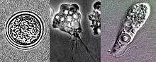
| Lifecycle stages of Naegleria fowleri: flagellate, trophozoite and cyst (seen from upper left to lower left to right) | |
| Scientific classification | |
| Phylum: | Percolozoa |
| Class: | Heterolobosea |
| Order: | Schizopyrenida |
| Family: | Vahlkampfiidae |
| Genus: | Naegleria |
| Species: | N. fowleri |
| Binomial name | |
| Naegleria fowleri Carter (1970) | |
Etymology
The organism was named after Dr. Malcolm Fowler, an Australian pathologist at Adelaide Children’s Hospital, who was the first author of the original series of case reports of primary amoebic meningoencephalitis.[4][5]
Life cycle
Naegleria fowleri is a thermophilic (heat-loving), free-living amoeba. It is found in warm and hot freshwater ponds, lakes and rivers, and in the very warm water of hot springs.[6] As the water temperature rises, its numbers increase. The amoeba was identified in the 1960s in Australia but appears to have evolved in the United States.[7] N. fowleri occurs in three forms – as a cyst, a trophozoite (ameboid), and a biflagellate (it has two flagella). It does not form a cyst in human tissue, where only the amoeboid trophozoite stage exists. The flagellate form can exist in the cerebrospinal fluid.
Cyst stage
The cyst form is spherical and about 7–15 µm in diameter. They are smooth, and have a single-layered wall with a single nucleus. Cysts are naturally resistant to environmental factors, so as to increase the chances of survival until better conditions occur. Trophozoites encyst due to unfavorable conditions. Factors that induce cyst formation include a lack of food, overcrowding, desiccation, accumulation of waste products, and cold temperatures.[8] When conditions improve, the amoeba can escape through the pore, or ostiole, seen in the middle of the cyst. N. fowleri has been found to encyst at temperatures below 10 °C (50 °F).[9]
Trophozoite stage
The trophozoite is the feeding, dividing, and infective stage for humans. The trophozoite attaches to olfactory epithelium, where it follows the olfactory cell axon through the cribriform plate (in the nasal cavity) to the brain. This reproductive stage of the protozoan organism, which transforms near 25 °C (77 °F) and grows best around 42 °C (106.7 °F), proliferates by binary fission. The trophozoites are characterized by a nucleus and a surrounding halo. They travel by pseudopodia, which means that they extend parts of their body's cell membrane (the pseudopods) and then fill them with plasma to force locomotion. The pseudopods form at different points along the cell, thus allowing the trophozoite to change directions. In their free-living state, trophozoites feed on bacteria. In tissues, it appears they phagocytize (consume by enclosing and then digesting prey) red blood cells and destroy tissue by releasing cytolytic substances.[8]
Flagellate
The flagellate is pear-shaped and biflagellate: this means that it has two flagella. This stage can be inhaled into the nasal cavity during swimming or diving. This biflagellate form occurs when trophozoites are exposed to a change in ionic concentration, such as placement in distilled water. The flagellate form does not exist in human tissue, but can exist in the cerebrospinal fluid. Once inside the nasal cavity, the flagellated form transforms into a trophozoite. The transformation of flagellate to trophozoite occurs within a few hours.[8]
Ecology
Naegleria fowleri are excavates that inhabit soil and water. N. fowleri is sensitive to drying and acid. It cannot survive in sea water. This amoeba is able to grow best at moderately elevated temperatures making summer month cases more likely. N. fowleri is somewhat of a thermophile and is able to grow at temperatures up to 46 °C (115 °F).[10] Warm, fresh water with a sufficient supply of bacterial food provides a habitat for amoebae. Man-made bodies of water, disturbed natural habitats, or areas with soil and unchlorinated/unfiltered water are locations where many amoebic infections have occurred.
N. fowleri seems to thrive during periods of disturbance; the flagellate-empty hypothesis explains that Nagleria's success may be due to decreased competition from a depleted population of the normal, thermosensitive protozoal fauna. In other words, N. fowleri thrives in the absence of other predators consuming its food supply. This hypothesis suggests that human disturbances such as thermal pollution increase N. fowleri abundance by removing their resource competitors. Ameoboflagellates have a motile flagellate stage that is designed for dispersal, which is advantageous when an environment has been cleared of competing organisms.
Pathogenicity
N. fowleri can cause an often lethal infection of the brain called naegleriasis (also known as primary amoebic meningoencephalitis, amoebic encephalitis/meningitis, or simply Naegleria infection). Infections most often occur when water containing N. fowleri is inhaled through the nose, where it then enters the nasal and olfactory nerve tissue, traveling to the brain through the cribriform plate.[11] N. fowleri normally eat bacteria, but during human infections, the trophozoites consume astrocytes and neurons. The reason why N. fowleri prefers to pass across the cribriform plate has remained unknown, but the neurotransmitter acetylcholine has been suggested to act as a stimulus, as a structural homolog of animal CHRM1 has been shown to be present in Naegleria and Acanthamoeba.[12]
It takes 1–9 days (average 5) for symptoms to appear after nasal exposure to N. fowleri flagellates.[13] Symptoms may include headache, fever and nausea. Later symptoms can include stiff neck, confusion, lack of attention, loss of balance, seizures, and hallucinations. Once symptoms begin to appear, death will usually occur within two weeks. A person infected with N. fowleri cannot spread the infection to another person.
While it is very rare, animals are in fact susceptible to infection by Naegleria fowleri. Experimentally, mice, guinea pigs, and sheep have been infected, and there have been cases reported where South American tapirs and cattle have contracted naegleriasis. The rarity of animal infection could be due to cases going largely unreported, however, as it stands animals can be infected by this amoeba. [14]
Treatment
The core antimicrobial treatment consists of antifungal drug amphotericin B,[15] which inhibits the pathogen by binding to its cell membrane sterols, thus leading to cell membrane disruption and pathogen death,[16] however the fatality rate even with this treatment is greater than 95%.[17] New treatments are being sought.[18] Miltefosine, an antiparasitic drug which inhibits the pathogen via disrupting its cell survival signal pathway PI3K/Akt/mTOR,[19] has been used in a few cases with mixed results.[20]
See also
- Acanthamoeba – an amoeba that can cause amoebic keratitis and encephalitis in humans
- Balamuthia mandrillaris – an amoeba that is the cause of (often fatal) granulomatous amoebic meningoencephalitis
- Entamoeba histolytica – an amoeba that is the cause of amoebiasis, or amoebic dysentery
- Leptospira – a zoonotic bacteria that causes leptospirosis
- Methicillin-resistant Staphylococcus aureus (MRSA)
- Necrotizing fasciitis – the "flesh-eating disease", caused by certain types of bacteria
- Toxoplasma gondii – pet-carried protozoan that causes the disease toxoplasmosis
- Vibrio vulnificus – warm saltwater infectious bacteria
References
- This happens to also be the common name of Balamuthia mandrillaris – An unrelated and even deadlier protist neuropathogen.
- Schuster, Frederick L., and Govinda S. Visvesvara. "Free-living Amoebae as Opportunistic and Non-opportunistic Pathogens of Humans and Animals." International Journal for Parasitology 34.9 (2004): 1001–1027. Web.
- "General Information: Naegleria fowleri". Centers for Disease Control and Prevention (CDC). Retrieved 2015-12-14.
- Fowler, M.; Carter, R.F. (September 25, 1965). "Acute Pyogenic Meningitis Probably due to Acanthamoeba sp.: a Preliminary Report" (PDF). British Medical Journal. 5464 (2): 740–742. doi:10.1136/bmj.2.5464.734-a. PMID 5825411. Retrieved August 15, 2019.
- "The discovery of amoebic meningitis in Northern Spencer Gulf towns". samhs.org. South Australian Medical Heritage Society Inc. Retrieved August 15, 2019.
- Laseke I, Korte J, Lamendella R, Kaneshiro ES, Marciano-Cabral F, Oerther DB (January 2010). "Identification of Naegleria fowler in warm ground water aquifers". Journal of Environmental Quality. 39 (1): 147–153. doi:10.2134/jeq2009.0062. PMID 20048302.
- "Brain-eating-amoeba". WebMD. Retrieved 1 July 2015.
- Marciano-Cabral, F (1988). "Biology of Naegleria spp". Microbiological Reviews. 52 (1): 114–133. PMC 372708. PMID 3280964.
- Chang, SL (1978). "Resistance of pathogenic Naegleria to some common physical and chemical agents". Applied and Environmental Microbiology. 35 (2): 368–375. PMC 242840. PMID 637538.
- "General Information | Naegleria fowleri | CDC". www.cdc.gov. 2018-07-17. Retrieved 2018-09-13.
- Baig, AM (Aug 2015). "Pathogenesis of amoebic encephalitis: Are the amoebae being credited to an 'inside job' done by the host immune response?". Acta Trop. 148: 72–76. doi:10.1016/j.actatropica.2015.04.022. PMID 25930186.
- Baig AM. Primary Amoebic Meningoencephalitis: Neurochemotaxis and Neurotropic Preferences of Naegleria fowleri. ACS Chem Neurosci. 2016 Aug 17;7(8):1026–1029. doi:10.1021/acschemneuro.6b00197. Epub 2016 July 22. PubMed PMID 27447543.
- "Naegleria fowleri – Primary Amebic Meningoencephalitis (PAM) – Amebic Encephalitis: Illness & Symptoms". Centers for Disease Control and Prevention (CDC).
- Naegleria Fowleri in Animals. Louisiana Dept of Health & Hospitals, 25 September 2013.
- Subhash Chandra Parija (Nov 23, 2015). "Naegleria Infection Treatment & Management". Medscape.
- Asbill, Scott, and Kris Virga. “Naegleria Fowleri: Pathogenesis, Diagnosis, and Treatment Options.” Antimicrobial Agents and Chemotherapy, American Society for Microbiology Journals, 1 Nov. 2015, aac.asm.org/content/59/11/6677.
- Cetin N, Blackall D. Naegleria fowleri meningoencephalitis. Blood. 2012 April 19;119(16):3658.PMID 22645743
- https://www.statnews.com/2016/07/22/brain-eating-amoeba/
- Asbill, Scott, and Kris Virga. “Naegleria Fowleri: Pathogenesis, Diagnosis, and Treatment Options.” Antimicrobial Agents and Chemotherapy, American Society for Microbiology Journals, 1 November. 2015, aac.asm.org/content/59/11/6677.
- Wessel, Linda (16 September 2016). "A life-saving drug that treats a rare infection is almost impossible to find". Business Insider. Archived from the original on 19 September 2016.
External links
| Wikimedia Commons has media related to Naegleria fowleri. |
- Naegleria information site from the Centers for Disease Control and Prevention
- Naegleria from The Tree of Life Web Project
- Naegleria fowleri at Stanford University's ParaSITE 2004