Superior longitudinal fasciculus
The superior longitudinal fasciculus (SLF) is an association fiber tract in the brain that is composed of three separate components.[1][2] It is present in both hemispheres and can be found lateral to the centrum ovale and connects the frontal, occipital, parietal, and temporal lobes.[2] These bundles of axon tracts pass from the frontal lobe through the operculum to the posterior end of the lateral sulcus where they either radiate to and synapse on neurons in the occipital lobe or turn downward and forward around the putamen and then radiate to and synapse on neurons in anterior portions of the temporal lobe.
| Superior longitudinal fasciculus | |
|---|---|
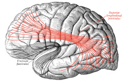 Lateral surface of left cerebral hemisphere. Some of major association tracts are depicted. Superior longitudinal fasciculus is at center, in red. | |
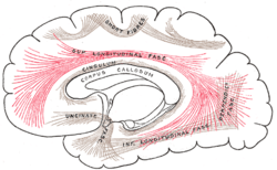 Diagram showing principal systems of association fibers in the cerebrum. (Sup. longitudinal fasc. labeled at center top.) | |
| Details | |
| Identifiers | |
| Latin | fasciculus longitudinalis superior cerebri |
| NeuroNames | 2080 |
| TA | A14.1.09.557 |
| FMA | 77631 |
| Anatomical terms of neuroanatomy | |
The SLF is composed of three distinct components SLF I, SLF II, and SLF III.[2][3]
SLF I
SLF I is the dorsal component and originates in the superior and medial parietal cortex, passes around the cingulate sulcus and in the superior parietal and frontal white matter, and terminates in the dorsal and medial cortex of the frontal lobe (Brodmann 6, 8, and 9) and in the supplementary motor cortex (M II).[4][5]
SLF I connects to the superior parietal cortex which encodes locations of body parts in a body-centric coordinate system and with M II and dorsal premotor cortex.[6] This suggests the SLF I is involved with regulating motor behavior, especially conditional associative tasks which select among competing motor tasks based on conditional rules.
SLF II
SLF II is the major component of SLF and originates in the caudal-inferior parietal cortex and terminates in the dorsolateral prefrontal cortex (Brodmann 6, 8 and 46).
SLF II connects to the caudal inferior parietal cortex which controls spatial attention and visual and oculomotor functions. This suggests the SLF II provides the prefrontal cortex with parietal cortex information regarding perception of visual space. Since these bundles are bi-directional, working memory (Brodmann 46) in the prefrontal cortex may provide the parietal cortex with information to focus spatial attention and regulate selection and retrieval of spatial information.
SLF III
SLF III is the ventral component and originates in the supramarginal gyrus (rostral portion of the inferior parietal lobe) and terminates in the ventral premotor and prefrontal cortex (Brodmann 6, 44, and 46).
SLF III connects the rostral inferior parietal cortex which receives information from the ventral precentral gyrus. This suggests that the SLF III transfers somatosensory information, such as language articulation, between the ventral premotor cortex, Brodmann 44 (pars opercularis), the supramarginal gyrus (Brodmann 40), and the laterial inferior prefrontal cortex working memory (Brodmann 46).
References
- Makris, N.; Kennedy, D. N.; McInerney, S.; Sorensen, A. G.; Wang, R.; Caviness, V. S.; Pandya, D. N. (2005-06-01). "Segmentation of Subcomponents within the Superior Longitudinal Fascicle in Humans: A Quantitative, In Vivo, DT-MRI Study". Cerebral Cortex. 15 (6): 854–869. doi:10.1093/cercor/bhh186. ISSN 1047-3211. PMID 15590909.
- Wang, Xuhui; Pathak, Sudhir; Stefaneanu, Lucia; Yeh, Fang-Cheng; Li, Shiting; Fernandez-Miranda, Juan C. (2016-05-01). "Subcomponents and connectivity of the superior longitudinal fasciculus in the human brain". Brain Structure & Function. 221 (4): 2075–2092. doi:10.1007/s00429-015-1028-5. ISSN 1863-2661. PMID 25782434.
- Makris N, et al. (2005). "Segmentation of subcomponents within the superior longitudinal fascicle in humans: a quantitative, in vivo, DT-MRI study". Cereb. Cortex. 15 (6): 854–55. doi:10.1093/cercor/bhh186. PMID 15590909.
- Kamali, A; Flanders, AE; Brody, J; Hunter, JV; Hasan, KM (2014). "Tracing superior longitudinal fasciculus connectivity in the human brain using high resolution diffusion tensor tractography". Brain Struct Funct. 219: 269–81. doi:10.1007/s00429-012-0498-y. PMC 3633629. PMID 23288254.
- Makris N, et al. (2005). "Segmentation of subcomponents within the superior longitudinal fascicle in humans: a quantitative, in vivo, DT-MRI study". Cereb. Cortex. 15 (6): 865. doi:10.1093/cercor/bhh186. PMID 15590909.
- Makris N, et al. (2005). "Segmentation of subcomponents within the superior longitudinal fascicle in humans: a quantitative, in vivo, DT-MRI study". Cereb. Cortex. 15 (6): 867. doi:10.1093/cercor/bhh186. PMID 15590909.
External links
| Wikimedia Commons has media related to Superior longitudinal fasciculus. |
- Brainmind.com atlas, see "1. Short arcuate bundles" section. Bundle 2 is the superior longitudinal fasciculus
- brain atlas sagittal images showing the SLF (bundle 2)
- Atlas image: n1a5p6 at the University of Michigan Health System - "Dissection of the Left Hemisphere"
| Authority control |
|---|