Inferior longitudinal fasciculus
The inferior longitudinal fasciculus is traditionally considered one of the major occipitotemporal association tracts. It connects the anterior temporal lobe and the extrastriate cortex of the occipital lobe, running along the lateral and inferior wall of the lateral ventricle.
| Inferior longitudinal fasciculus | |
|---|---|
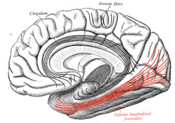 Medial surface of right cerebral hemisphere. Some of major association tracts are depicted. Inferior longitudinal fasciculus labeled at bottom right, in red. | |
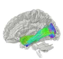 Tractography showing inferior longitudinal fasciculus | |
| Details | |
| Identifiers | |
| Latin | fasciculus longitudinalis inferior cerebri |
| NeuroNames | 1443 |
| TA | A14.1.09.556 |
| FMA | 77632 |
| Anatomical terms of neuroanatomy | |
The existence of this fasciculus and its anatomical description[1][2] have been the subject of several mutually conflicting studies. Some authors denied its existence because of the unclear results obtained in non-human brains.[3][4]
Using diffusion tensor imaging (DTI), several authors have confirmed the presence of this constant longitudinal pathway in humans.[5][6][7][8][9]
Some other studies of the ILF[10][11][12][13] based on Klingler's dissection method (a type of white matter blunt dissection, providing reliable data on the anatomy of major fibre bundles[14] and, in some cases, additional tractography[13][15] not only confirmed the classical descriptions of the direct connection between occipital and temporal regions but also sought to detail the subcomponents of this association tract.
Four branches were consistently identified: a fusiform branch connecting the fusiform gyrus to the anterior temporal regions; a dorsolateral occipital branch connecting the superior, middle and inferior occipital gyri to the anterior temporal regions; a lingual branch connecting the lingual gyrus to the anterior part of the middle temporal gyrus; and a minor cuneal branch connecting the cuneus to the anterior mesial temporal gyri.[13][15]
Functions of the ILF
Summarising studies from healthy individuals, intraoperative and lesional findings, this white matter bundle seems involved in a wide range of brain conditions, including psychopathological, neurodevelopmental and neurological diseases. Among these diseases and syndromes are i.e. associative visual agnosia,[16] prosopagnosia,[17][18] visual amnesia,[19] visual hypo-emotionality;[20][21][22][23] but also some forms of autism spectrum disorders, schizophrenia and alexia.[24][25][26]
ILF supports brain functions concerning the visual modality, including object, face and place processing, reading, lexical and semantic processing, emotion processing, and visual memory. Based on these recent findings ILF can be described as a multi-functional white matter pathway involved in visually guided behavior (See Herbet et al. for review[27]).
References
- Dejerine, J., and Dejerine-Klumpke, A. (1895). Anatomie des centres nerveux. Rueff.
- Polyak, S., 1957. The vertebrate visual system. Chicago: University of Chicago Press
- Putnam, T.J., 1926. Studies on the central visual connections. Arch Neurol Psychiatr; 16: 566±96
- Tusa, R.J., Ungerleider, L.G., 1985. The inferior longitudinal fasciculus: a reexamination in humans and monkeys. Ann Neurol; 18: 583-91.
- Mori, S., Kaufmann, W.E., Davatzikos, C., Stieltjes, B., Amodei, L., Fredericksen, K., Pearlson, G.D., Melhem, E.R., Solaiyappan, M., Raymond, G.V., Moser, H.W., van Zijl, P.C. 2002. Imaging cortical association tracts in the human brain using diffusion-tensor-based axonal tracking. MagnResonMed.;47(2):215-23
- Catani, M., Howard, R.J., Pajevic, S., Jones, D.K. 2002. Virtual in vivo interactive dissection of white matter fasciculi in the human brain. Neuroimage;17(1):77-94
- Catani, M., Jones, D.K., Donato, R., Ffytche, D.H., 2003. Occipito-temporal connections in the human brain. Brain; 126: 2093-107.
- Wakana, S., Caprihan, A., Panzenboeck, M.M., Fallon, J.H., Perry, M., Gollub, R.L., Hua, K., Zhang, J., Jiang, H., Dubey, P., Blitz, A., van Zijl, P., Mori, S. 2007. Reproducibility of quantitative tractography methods applied to cerebral white matter. Neuroimage; 1;36(3):630-44.
- Catani, M., Thiebaut de Schotten, M., 2008. A diffusion tensor imaging tractography atlas for virtual in vivo dissections. Cortex.;44(8):1105-32.
- Fernández-Miranda, J.C., Rhoton, A.L., Álvarez-Linera, J., Kakizawa, Y., Choi, C., De Oliveira, E., 2008. Three Dimensional Microsurgical and Tractographic anatomy of the white matter of the Human Brain. Neurosurgery 62[SHC Suppl 3]: SHC-989-SHC-1027.
- De Benedictis, A., Duffau, H., Paradiso, B., Grandi, E., Balbi, S., Granieri, E., Colarusso, E., Chioffi, F., Marras, C.E., Sarubbo, S., 2014. Anatomo-functional study of the temporo-parieto-occipital region: dissection, tractographic and brain mapping evidence from a neurosurgical perspective. J Anat;225(2):132-51
- Latini, F. (2015). New insights in the limbic modulation of visual inputs: the role of the inferior longitudinal fasciculus and the Li-Am bundle. Neurosurgical Review 38, 179– 190
- Latini, F., Mårtensson, J., Larsson, E.-M., Fredrikson, M., Åhs, F., Hjortberg, M., et al. (2017). Segmentation of the inferior longitudinal fasciculus in the human brain: A white matter dissection and diffusion tensor tractography study. Brain Research
- Agrawal, A., Kapfhammer, J.P., Kress, A., Wichers, H., Deep, A., Feindel, W., Sonntag, V.K., Spetzler, R.F., Preul, M.C., 2011. Josef Klingler’s models of white matter tracts: influences on neuroanatomy, neurosurgery, and neuroimaging. Neuro- surgery 69, 238–252
- Panesar SS, Yeh FC, Jacquesson T, Hula W, Fernandez-Miranda JC. A Quantitative Tractography Study Into the Connectivity, Segmentation and Laterality of the Human Inferior Longitudinal Fasciculus.Front Neuroanat. 2018 Jun 5;12:47. doi: 10.3389/fnana.2018.00047
- Jankowiak, J., Albert, M.L., 1994. Lesion localization in visual agnosia. In: Kertesz A, editor. Localization and neuroimaging in neuropsychology. San Diego: Academic Press; p. 429±71
- Benson, D.F., Segarra, J., Albert, M.L., 1974. Visual agnosia-prosopagnosia. A clinicopathologic correlation. Arch Neurol; 30: 307-10.
- Meadows, J.C., 1974. The anatomical basis of prosopagnosia. J Neurol Neurosurg Psychiatry; 37: 489-501
- Ross, E.D., 1980. Sensory-specific and fractional disorders of recent memory in man. I. Isolated loss of visual recent memory. Arch Neurol; 37: 193±200
- Bauer, R.M., 1982. Visual hypoemotionality as a symptom of visual±limbic disconnection in man. Arch Neurol; 39: 702-8.
- Sierra, M., Lopera, F., Lambert, M.V., Phillips, M.L., David, A.S., 2002. Separating depersonalisation and derealisation: the relevance of the `lesion method'. J Neurol Neurosurg Psychiatry; 72: 530-2.
- Geschwind, N., 1965a .Disconnexion syndromes in animals and man. Part I. Brain; 88: 237±94
- Girkin, C.A., Miller, N.R., 2001. Central disorders of vision in humans. Surv Ophthalmol; 45: 379±405
- Ashtari, M., Cottone, J., Ardekani, B.A., Cervellione, K., Szeszko, P.R., Wu, J., Chen, S., Kumra, S., 2007. Disruption of white matter integrity in the inferior longitudinal fasciculus in adolescents with schizophrenia as revealed by fiber tractography. Archives of General Psychiatry, 64(11): 1270e1280
- Epelbaum, S., Pinel, P., Gaillard, R., Delmaire, C., Perrin, M., Dupont, S., Dehaene, S., Cohen, L., 2008. Pure alexia as a disconnection syndrome: new diffusion imaging evidence for an old concept. Cortex. Sep;44(8):962-74. doi: 10.1016/j.cortex.2008.05.003
- Koldewyn, K., Yendiki, A., Weigelt, S., Gweon, H., Julian, J., Richardson, H., Malloy, C., Saxe, R., Fischl, B., Kanwisher, N., 2014. Differences in the right inferior longitudinal fasciculus but no general disruption of white matter tracts in children with autism spectrum disorder. Proc Natl Acad Sci U S A. Feb 4;111(5):1981-6. doi: 10.1073/pnas.1324037111
- Herbet G, Zemmoura I, Duffau H. Functional Anatomy of the Inferior Longitudinal Fasciculus: From Historical Reports to Current Hypotheses. Front Neuroanat. 2018 Sep 19;12:77. doi: 10.3389/fnana.2018.00077
External links
| Wikimedia Commons has media related to Inferior longitudinal fasciculus. |
- Atlas image: n1a5p6 at the University of Michigan Health System - "Dissection of the Left Hemisphere"
| Authority control |
|---|