Arcuate fasciculus
The arcuate fasciculus (Latin: curved bundle) is a bundle of axons that connects Broca's area and Wernicke's area in the brain. It is an association fiber tract connecting caudal temporal cortex and inferior frontal lobe.[1]
| Arcuate fasciculus | |
|---|---|
 The arcuate fasciculus connects two important areas for language use, Broca's area and Wernicke's area. | |
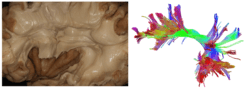
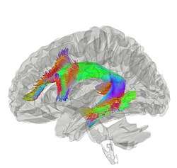 Tractography showing arcuate fasciculus | |
| Details | |
| From | Broca's area of the frontal lobe |
| To | Wernicke's area of the temporal lobe |
| Identifiers | |
| Latin | fasciculus arcuatus |
| TA | A14.1.09.557 |
| FMA | 260714 |
| Anatomical terms of neuroanatomy | |
Structure
Common understanding has been that the arcuate fasciculus connects two important areas for language use, Broca's area in the inferior frontal gyrus and Wernicke's area in the posterior superior temporal gyrus. The connectivity of the arcuate has been shown to correspond to various functional areas within the temporal, parietal, and frontal lobes.[2] Furthermore, the topographical relationships between independent measures of white matter and gray matter integrity suggest that rich developmental or environmental interactions influence brain structure and function, and that the presence and strength of such associations may elucidate pathophysiological processes influencing systems such as language and motor planning.
As the technique of diffusion MRI has improved, this has become a testable hypothesis using brain imaging. Research indicates more diffuse termination of the fibers of the arcuate, both rostrally and caudally, than previously thought. While the main caudal source of the fiber tract appears to be posterior superior temporal cortex, the rostral terminations are mostly in premotor cortex, part of Brodmann area 6.[3][4][5]
Clinical significance
Evidence for the role of the arcuate fasciculus in language use is best represented by conduction aphasia, caused by damage to the inferior parietal lobule that extends into the subcortical white matter and damages the arcuate fasciculus.[6] This type of aphasia inhibits the patient from repeating unfamiliar sounds. Damage to the direct pathway may produce conduction aphasia, whereas damage to the indirect pathway spares the ability to repeat speech but impairs comprehension. The symptoms of conduction aphasia suggest that the connection between posterior temporal cortex and frontal cortex plays a vital role in short-term memory of words and speech sounds that are new or have just been heard. The arcuate fasciculus connects these two regions and circulates information back and forth, possibly contributing to short-term memory.
In nine out of ten people with tone deafness, the superior arcuate fasciculus in the right hemisphere could not be detected, suggesting a disconnection between the posterior superior temporal gyrus and the posterior inferior frontal gyrus. Researchers suggested the posterior superior temporal gyrus was the origin of the disorder.[7]
In stutterers, the arcuate fasciculus appears to have bilateral deficits that reduce it by one-third or more relative to non-stutterers.[8] However, there is ongoing debate concerning the contribution of each hemisphere and the presence of hemispheric differences, and diffusion-based evidence of differences between stutterers and controls is not isolated to the arcuate fasciculus.
See also
- Aphasia
- Wernicke–Geschwind model
References
- Carlson, N. (2012). Physiology of behavior. (11th ed.). Pearson.
- Phillips, Owen (2011). "Topographical relationships between arcuate fasciculus connectivity and cortical thickness". Research. Human Brain Mapping. 32 (11): 1788–1801. doi:10.1002/hbm.21147. PMC 3071430. PMID 20886580.
- CATANI, M; THIEBAUT DE SCHOTTEN, M (1 September 2008). "A diffusion tensor imaging tractography atlas for virtual in vivo dissections". Cortex. 44 (8): 1105–1132. doi:10.1016/j.cortex.2008.05.004.
- Bernal, B.; Ardila, A. (18 August 2009). "The role of the arcuate fasciculus in conduction aphasia". Brain. 132 (9): 2309–2316. doi:10.1093/brain/awp206. PMID 19690094. Retrieved 5 October 2013.
- Bernal, Byron; Altman, Nolan (1 February 2010). "The connectivity of the superior longitudinal fasciculus: a tractography DTI study". Magnetic Resonance Imaging. 28 (2): 217–225. doi:10.1016/j.mri.2009.07.008. PMID 19695825.
- Adams, R. D. The anatomy of memory mechanisms in the human brain. In "The pathology of Memory, edited by G. A. Talland and N. C. Waugh. New York: Academic Press, 1969.
- Loui, P; Alsop, D; Schlaug, S (2009). "Tone Deafness: A New Disconnection Syndrome?". Journal of Neuroscience. 29 (33): 10215–10220. doi:10.1523/JNEUROSCI.1701-09.2009. PMC 2747525. PMID 19692596.
- Cieslak, M; Ingham, R; Ingham, J; Grafton, S (2015). "Anomalous white matter morphology in adults who stutter". Journal of Speech, Language, and Hearing Research. 58 (2): 268–277. doi:10.1044/2015_JSLHR-S-14-0193. PMC 4675119. PMID 25635376.
External links
| Wikimedia Commons has media related to Arcuate fasciculus. |
- https://web.archive.org/web/20070528184714/http://www.lib.mcg.edu/edu/eshuphysio/program/section8/8ch15/s8c15_14.htm
- http://thebrain.mcgill.ca/flash/d/d_10/d_10_cr/d_10_cr_lan/d_10_cr_lan.html
- http://brain.oxfordjournals.org/cgi/reprint/awh622v1.pdf
| Authority control |
|---|