Phycobilisome
Phycobilisomes are light harvesting antennae of photosystem II in cyanobacteria, red algae and glaucophytes.
| Phycobilisome protein | |||||||||
|---|---|---|---|---|---|---|---|---|---|
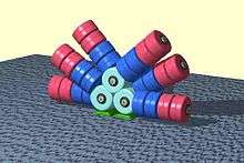 The layout of protein subunits in a phycobilisome. | |||||||||
| Identifiers | |||||||||
| Symbol | Phycobilisome | ||||||||
| Pfam | PF00502 | ||||||||
| InterPro | IPR012128 | ||||||||
| SCOPe | 1cpc / SUPFAM | ||||||||
| |||||||||
General structure
Phycobilisomes are protein complexes (up to 600 polypeptides) anchored to thylakoid membranes. They are made of stacks of chromophorylated proteins, the phycobiliproteins, and their associated linker polypeptides. Each phycobilisome consists of a core made of allophycocyanin, from which several outwardly oriented rods made of stacked disks of phycocyanin and (if present) phycoerythrin(s) or phycoerythrocyanin. The spectral property of phycobiliproteins are mainly dictated by their prosthetic groups, which are linear tetrapyrroles known as phycobilins including phycocyanobilin, phycoerythrobilin, phycourobilin and phycobiliviolin. The spectral properties of a given phycobilin is influenced by its protein environment.[1]
Function
Each phycobiliprotein has a specific absorption and fluorescence emission maximum in the visible range of light. Therefore, their presence and the particular arrangement within the phycobilisomes allow absorption and unidirectional transfer of light energy to chlorophyll a of the photosystem II. In this way, the cells take advantage of the available wavelengths of light (in the 500-650 nm range), which are inaccessible to chlorophyll, and utilize their energy for photosynthesis. This is particularly advantageous deeper in the water column, where light with longer wavelengths is less transmitted and therefore less available directly to chlorophyll.
The geometrical arrangement of a phycobilisome is very elegant in a antenna-like assembly. It results in 95% efficiency of energy transfer.[2]
Evolution and diversity
There are many variations to the general phycobilisomes structure. Their shape can be hemidiscoidal (in cyanobacteria) or hemiellipsoidal (in red algae). Species lacking phycoerythrin have at least two disks of phycocyanin per rod, which is sufficient for maximum photosynthesis.[3]
The phycobiliproteins themselves show little sequence evolution due to their highly constrained function (absorption and transfer of specific wavelengths). In some species of cyanobacteria, when both phycocyanin and phycoerythrin is present, the phycobilisome can undergo significant restructuring as response to light color. In green light the distal portions of the rods are made of red colored phycoerythrin, which absorbs green light better. In red light, this is replaced by blue colored phycocyanin, which absorbs red light better. This reversible process is known as complementary chromatic adaptation. It is the component of photosynthetic system of cyanobacteria, as a particle with which various structures are linked (i.e. thylakoid membrane, etc).
Applications
Phycobilisomes can be used in prompt fluorescence,[4][5] flow cytometry,[6] Western blotting and protein microarrays. Some phycobilisomes have an absorption and emission profile similar to Cy5, they can be used in many of the same applications, however, they can be up to 200 times brighter, with a large Stokes shift, providing a larger signal per binding event. This property allows the detection of low-level target molecules]\[6] or rare events.
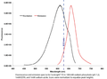 Excitation and emission spectra of a phycobilisome from a blue-green alga.
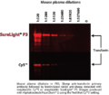 Phycobilisome versus cyanine dye detection capabilities in Western blot application.
References
- Singh, NK; Sonani, RR; Rastogi, RP; Madamwar, D (2015). "The phycobilisomes: an early requisite for efficient photosynthesis in cyanobacteria". EXCLI journal. 14: 268–89. doi:10.17179/excli2014-723. PMC 4553884. PMID 26417362.
- Light Harvesting by Phycobilisomes Annual Review of Biophysics and Biophysical Chemistry Vol. 14: 47-77 (Volume publication date June 1985)
- Lea-Smith DJ, Bombelli P, Dennis JS, Scott SA, Smith AG, Howe CJ (June 2014). "Phycobilisome-Deficient Strains of Synechocystis sp. PCC 6803 Have Reduced Size and Require Carbon-Limiting Conditions to Exhibit Enhanced Productivity". Plant Physiology. 165 (2): 705–714. doi:10.1104/pp.114.237206. PMC 4044857. PMID 24760817.
- Zoha SJ, Ramnarain S, Morseman JP, Moss MW, Allnutt FC, Rogers Y, Harvey B (1999). "PBXL Fluorescent Dyes for Ultrasensitive Direct Detection". Journal of Fluorescence. 9 (3): 197–208. doi:10.1023/A:1022503600141.
- "MicroPlate Detection comparison between SureLight®P-3L, other fluorophores and enzymatic detection" (PDF). Columbia Biosciences. 2010.
- Telford WG, Moss MW, Morseman JP, Allnutt FC (August 2001). "Cyanobacterial stabilized phycobilisomes as fluorochromes for extracellular antigen detection by flow cytometry" (PDF). Journal of Immunological Methods. 254 (1–2): 13–30. doi:10.1016/s0022-1759(01)00367-2. PMID 11406150.
External links
- Phycobilisomes at the US National Library of Medicine Medical Subject Headings (MeSH)
- Columbia Biosciences - Phycobilisome Resource