Frontonasal process
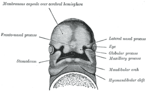
The frontonasal process, or frontonasal prominence is one of the five swellings that develop to form the face. The frontonasal process is unpaired, and the others are the paired maxillary prominences, and the paired mandibular prominences. During the fourth week of embryonic development, an area of thickened ectoderm develops, on each side of the frontonasal process called the nasal placodes or olfactory placodes, and appear immediately under the forebrain.[1]
| Frontonasal process | |
|---|---|
 Under surface of the head of a human embryo about twenty-nine days old. (Frontonasal process labeled at center left.) | |
| Details | |
| Precursor | Ectoderm |
| Identifiers | |
| TE | E5.3.0.0.0.0.6 |
| Anatomical terminology | |
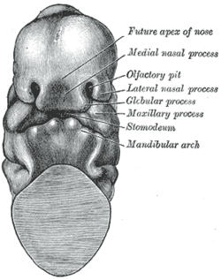
By invagination these areas are converted into two nasal pits, which indent the frontonasal prominence and divide it into medial and lateral nasal processes.[2]
Nasal processes

The medial nasal process (nasomedial) on the inner side of each nasal pit merge into the intermaxillary segment and form the upper lip, crest, and tip of the nose.[1] The medial nasal processes merge with the maxillary prominences. The lateral nasal process from each side merge to form the alae of the nose.[1]
Genetics
There is some evidence that development involves Sonic hedgehog and Fibroblast growth factor 8.[3]
References
This article incorporates text in the public domain from page 67 of the 20th edition of Gray's Anatomy (1918)
- Sadler, T (2006). Langman's Medical Embryology. pp. 280–284. ISBN 9780781790697.
- Larsen, W (2001). Human embryology. pp. 365–368. ISBN 0443065837.
- Abzhanov A, Cordero DR, Sen J, Tabin CJ, Helms JA (December 2007). "Cross-regulatory interactions between Fgf8 and Shh in the avian frontonasal prominence". Congenit Anom (Kyoto). 47 (4): 136–48. doi:10.1111/j.1741-4520.2007.00162.x. PMID 17988255.
External links
- hednk-027—Embryo Images at University of North Carolina
- Flash animation at indiana.edu
- ent/30 at eMedicine